The Basics of ECG
The information contained within a single 12-lead electrocardiogram can be extensive. Learning how to interpret the subtle differences in characteristic changes that can arise is a specialized skill that can take years to learn. Fortunately, basic ECG interpretation can be rather straightforward, as long as you know the basics.
An electrocardiogram is a tracing of the electrical activity that is taking place within the heart. Under normal circumstances, an electrical impulse will travel from the sinoatrial node, spread across the atrium, to the atrioventricular node and through the ventricular septum of the heart. This electrical impulse causes the four chambers of the heart to contract and relax in a coordinated fashion. Studying these electrical impulses allows us to understand how the heart is functioning.
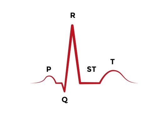
P Wave
The P wave represents the depolarization of the left and right atrium and also corresponds to atrial contraction. Strictly speaking, the atria contract a split second after the P wave begins. Because it is so small, atrial repolarization is usually not visible on ECG. In most cases, the P wave will be smooth and rounded, no more than 2.5 mm tall, and no more than 0.11 seconds in duration. It will be positive in leads I, II, aVF and V1 through V6.
QRS Complex
As the name suggests, the QRS complex includes the Q wave, R wave, and S wave. These three waves occur in rapid succession. The QRS complex represents the electrical impulse as it spreads through the ventricles and indicates ventricular depolarization. As with the P wave, the QRS complex starts just before ventricular contraction.
It is important to recognize that not every QRS complex will contain Q, R, and S waves. The convention is that the Q wave is always negative and that the R wave is the first positive wave of the complex. If the QRS complex only includes an upward (positive) deflection, then it is an R wave. The S wave is the first negative deflection after an R wave.
Under normal circumstances, the duration of the QRS complex in an adult patient will be between 0.06 and 0.10 seconds. The QRS complex is usually positive in leads I, aVL, V5, V6 and II, III, and aVF. The QRS complex is usually negative in leads aVR, V1, and V2.
The J-point is the point where the QRS complex and the ST segment meet. It can also be thought of as the start of the ST segment. The J-point (also known as Junction) is important because it can be used to diagnose an ST segment elevation myocardial infarction. When the J-point is elevated at least 2 mm above baseline, it is consistent with a STEMI.
T Wave
A T wave follows the QRS complex and indicates ventricular repolarization. Unlike a P wave, a normal T wave is slightly asymmetric; the peak of the wave is a little closer to its end than to its beginning. T waves are normally positive in leads I, II, and V2 through V6 and negative in aVR. A T wave will normally follow the same direction as the QRS complex that preceded it (positive or negative/up or down). When a T wave occurs in the opposite direction of the QRS complex, it generally reflects some sort of cardiac pathology.
If a small wave occurs between the T wave and the P wave, it could be a U wave. The biological basis for a U wave is unknown.
Heart Rate
There are many ways to determine a patient’s heart rate using ECG. One of the quickest ways is called the sequence method. To use the sequence method, find an R wave that lines up with one of the dark vertical lines on the ECG paper. If the next R wave appears on the next dark vertical line, it corresponds to heart rate of 300 beats a minute. The dark vertical lines correspond to 300, 150, 100, 75, 60, and 50 bpm. For example, if there are three large boxes between R waves, the patient’s heart rate is 100 bpm. There are more accurate ways to determine heart rate from ECG, but in life-saving scenarios, this method provides a quick estimate.
Mastering EKG interpretation is the foundation of accurate cardiac care. Sharpen your rhythm recognition skills and advance your expertise through our ACLS certification online program.
- 100% Online Training
- 3 Exam Attempts
- 2 Year Certification
- 100% Online Training
- 3 Exam Attempts
- 2 Year Certification
- 100% Online Training
- 3 Exam Attempts
- 2 Year Certification
- 100% Online Training
- 3 Exam Attempts
- 2 Year Certification
Pricing for New Customers Only
- Latest ECC & ILCOR Guidelines
- No Skills Test Required
- 24/7 Online Access
- Instant Card Access
- 2 Year Certification