Category Archives: Latest News

Unstable Bradycardia Resolves Following Atropine and Attempted Transcutaneous Pacing (TCP)

A  75 year old male experienced a syncopal episode. The event was witnessed by family members who contacted 9-1-1. On arrival of EMS personnel the patient appears acutely ill. He is pale, diaphoretic and cool to touch. He states that he is feeling lightheaded and weak.

Medical History

  • Hypertension
  • Hyperlipidemia
  • Gout
  • Bilateral knee replacement
  • Left bundle branch block

The family reports the patient is seeing a cardiologist and is scheduled for pacemaker implantation in 3 weeks due to previous episodes of symptomatic bradycardia.

Medications

  • Zocor (Simvastatin)
  • Lopressor (Metoprolol)
  • Aloprim (Allopurinol)
  • Multi-vitamins

Vital Signs

  • RR: 20
  • HR: 20
  • BP: 80/48 mm Hg
  • SpO2: 93% on room air
  • Capillary blood glucose: 118 mg/dL

Breath sounds are clear bilaterally.

The patient is placed on O2 via nasal cannula at 2 LPM with ETCO2 of 16 mmHg.

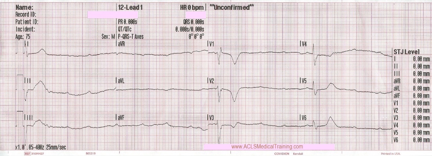
Cardiac monitoring is established and the following 12 lead ECG is obtained.

High degree AV block with heart rate less than 20.

When considering bradycardia management, 3 initial questions should be answered:

  • Is the patient bradycardic?
  • Is the patient symptomatic?
  • Is the patient symptomatic from the bradycardia?

If all of these have been answered with a YES, determine the patient’s hemodynamic status.

Signs of hemodynamically instability:

  • Altered mental status
  • Hypotension
  • Ischemic chest pain
  • Signs of hypoperfusion
  • Acute pulmonary edema

Based on these criteria this patient is clearly unstable.

Defibrillation pads are placed as a precaution, IV access is obtained, and 250 ml of normal saline is administered en route to the Emergency Department which is 4 minutes away from the scene.

Upon arrival blood samples are obtained and the following 12 lead ECG is obtained.
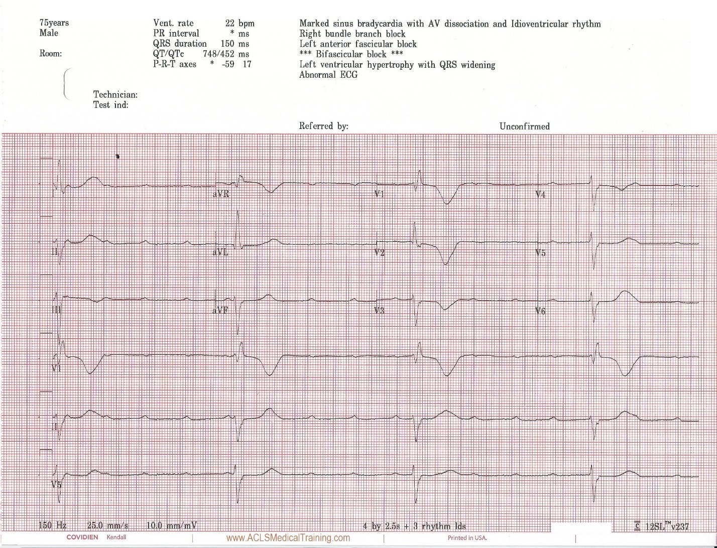
3rd Degree AV Block. The escape rhythm shows a wide QRS with bifascicular morphology (RBBB morphology with left axis deviation). It is likely a ventricular in origin.

The patient’s level of consciousness deteriorates and he responds only to painful stimuli.

0.5 mg atropine is administered rapid IV push followed by 10 ml saline flush.

After 1 minute transcutaneous pacing is initiated with no electrical capture up to 90 mA. Transcutaneous pacing is discontinued by the arriving cardiologist who requests vasopressors.

Prior to vasopressors being administered a change is noted on the cardiac monitor and another 12 lead ECG is obtained.
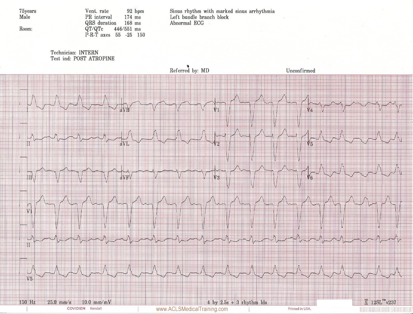
The heart rate is now 92. There is left bundle branch block which is consistent with the patient’s known medical history. At first glance this appears to be sinus rhythm although the last 3 cardiac cycles make the exact rhythm uncertain.

The patient now reports he is feeling better. His skin color improves and his blood pressure normalizes.

The patient was taken to cardiac cath lab for angiography and a permanent pacemaker. The procedure was successful and he was placed in the cardiac step-down unit for further observation.

Discussion

Atropine is an anticholinergic drug – also known as a parasympatholytic – which means that it counteracts increased vagal tone by binding to cardiac muscarinic receptors, which can improve sinus, atrial, and AV-nodal conduction. However, it is not believed to have a direct affect on rhythms of ventricular origin.

The 2010 AHA ECC Guidelines caution:

“Avoid relying on atropine in type II second-degree or third-degree AV block or in patients with third-degree AV block with a new wide-QRS complex where the location of block is likely to be in non-nodal tissue (such as in the bundle of His or more distal conduction system). These bradyarrhythmias are not likely to be responsive to reversal of cholinergic effects by atropine and are preferably treated with TCP…”

There is a caveat regarding TCP:

“TCP is, at best, a temporizing measure. TCP is painful in conscious patients, and, whether effective or not (achieving inconsistent capture), the patient should be prepared for transvenous pacing and expert consultation should be obtained. It is reasonable for healthcare providers to initiate TCP in unstable patients who do not respond to atropine. Immediate pacing might be considered in unstable patients with high-degree AV block when IV access is not available. If the patient does not respond to drugs or TCP, transvenous pacing is probably indicated.”

In this case transcutaneous pacing was not successful but the milliamperes were not increased beyond 90 milliamperes. On the plus side, the clinicians who were caring for this patient realized that they had not achieved capture, which is not always the case!

Fortunately, the patient spontaneously converted into a perfusing rhythm. The atropine may have contributed to the termination of this 3rd degree AV block, it may have been related to sympathetic stimulation from attempted transcutaneous pacing, or it could be a coincidence. These types of scenarios are susceptible to the “post hoc ergo propter hoc” fallacy (after this, therefore because of this).

A common error when treating patients with bradycardia is a rush to drug or electrical therapy prior to identifying reversible causes. Remember, Hs and Ts aren’t just for asystole and PEA!

Most importantly, hypoxemia should be rapidly identified and treated, but other conditions like hyperkalemia can also cause bradycardia. There is little to lose and much to gain from giving a patient calcium gluconate or calcium chloride prior to pacing. The quote Stephen Smith, M.D.: “The treatment [of hyperkalemia with calcium] is benign and cheap. How many life-threatening diseases can you treat benignly and cheaply?”

The definitive care for this patient was a permanent pacemaker.

Further reading: ACLS Bradycardia Algorithm

References

Neumar R, Otto C, Link M et al. Part 8: Adult Advanced Cardiovascular Life Support: 2010 American Heart Association Guidelines for Cardiopulmonary Resuscitation and Emergency Cardiovascular Care. Circulation. 2010;122(18_suppl_3):S729-S767. doi:10.1161/circulationaha.110.970988.

Abrich V, Le R, Friedman P, et al. Aggressive Management of Bradycardia is Associated With Improved Clinical Outcomes and Shorter Length of Stay: A Comparison of Two Academic Centers. Circulation. 2016;134:A20838

EMCrit Podcast 42: A phD in EKG with Steve Smith. EMCrit Podcast. 2011. Available at: https://emcrit.org/emcrit/phd-in-ekg/. Accessed November 26, 2017.

 

Mixed Overdose and Na-Channel Blockade

It’s 06:30 when EMS is called to an inner-city apartment for an 18-year-old male having a seizure. After gaining entry into the building, Paramedics and First Responders trudge up two flights of stairs and down a narrow, dimly lit hallway, until they find an open door into a dark two-bedroom apartment.  There are three people in the living room, all about 18-years-old, and one of them is lying awkwardly on the floor, propped up against the side of the couch. There is evidence of extensive alcohol and drug consumption littering the scene.

It proves difficult to obtain a complete patient history, since bystanders on scene are still somewhat inebriated, and they remain apprehensive about cooperating with emergency responders. However, the story gathered is as follows.

The three of them were up all night partying, drinking, and ingesting multiple illicit substances. It is reported that the patient was witnessed to have ingested copious amounts of cocaine, “DXM” (Dextromethorphan, or ‘cough syrup’), marijuana, nicotine, and alcohol. Precise amounts are vague, and it’s possible that there may have been even more ingestions that were not reported. The party ended, and they all went to sleep, until one of the party-goers found the patient seizing on the floor at about 06:30 this morning. It’s unknown how long the patient was down prior to being found.

The patient’s medical history is unknown, though it is believed that he is generally a “pretty healthy guy”.

The patient is unresponsive to any stimuli, has a weak and agonal respiratory effort, and a faint and slow carotid pulse. His general appearance is poor, with significant pallor and central cyanosis noted, though he is hot to the touch.

Crews began administering high-quality 2-person BVM ventilation, utilizing a jaw thrust while inserting an OPA, and positioning the patient with padding behind the head to support a proper ear-to-sternal-notch alignment. Intravenous access, fluid resuscitation, reoxygenation, and basic cardiac monitoring is being maintained while extrication from the apartment is coordinated.

His initial vital signs are as follows:

HR – 60/min, and increasing to 100/min once oxygenation and ventilation is administered
RR – 6/minute and ineffective
SpO2 Initially <50%, improving to 83% with oxygenation and ventilation
NIBP – 66/34 [44]
Pupils – 6mm bilaterally, extremely sluggish
BGL – 5.0 mmol/L (90 mg/dl)
Temp – 38.5 Celsius (101.3 F)

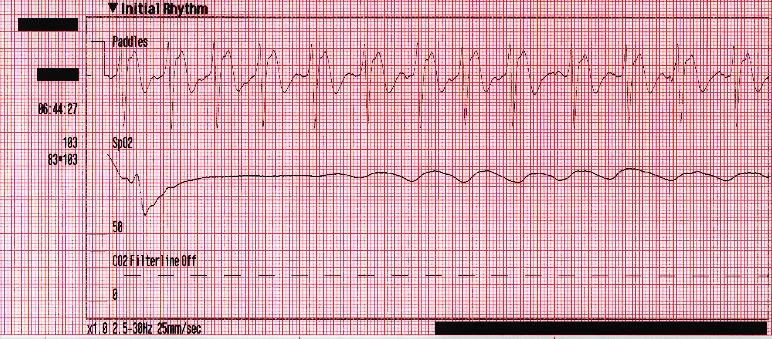
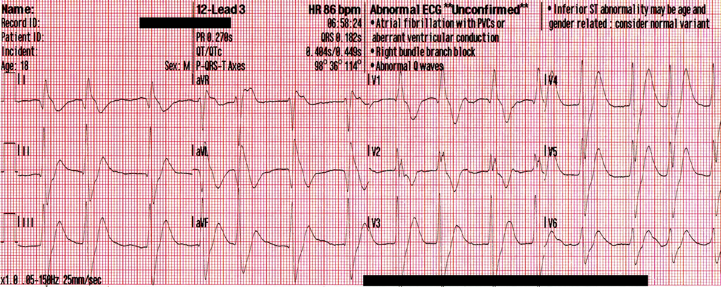
The following rhythm is observed on the monitor. Shortly later, the patient has a generalized seizure which lasts approximately 5 minutes.

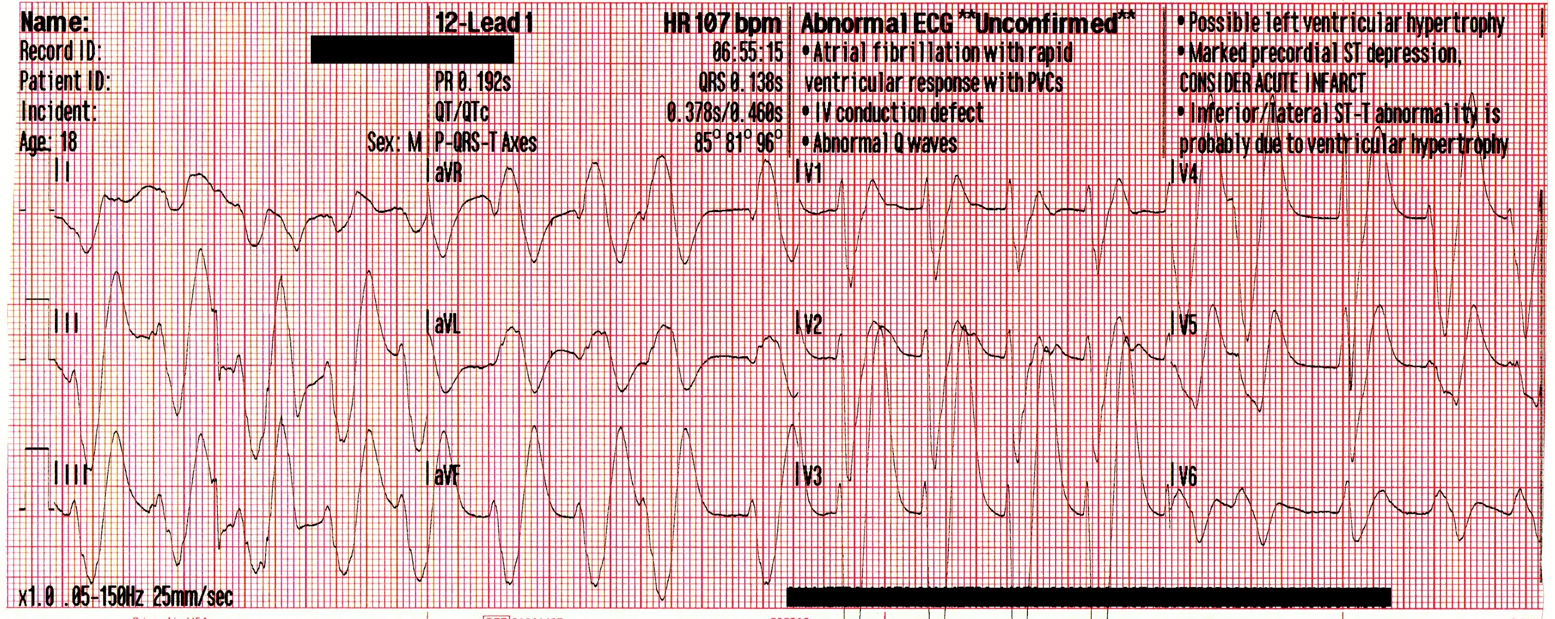
Due to access limitations, the stretcher could not be brought inside to the patient, and so the team of paramedics, police officers and firefighters worked together to move the patient to the ambulance via a device not unlike a large tarp with handles. Once in the ambulance, passive cooling is initiated, and a supraglottic airway is placed. His blood pressure has improved to 87/44 [59], his SpO2 remains in the mid-80’s, and his initial ETCO2 is 86mmHg. The following 12-lead is acquired:

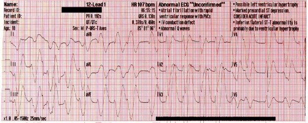
A significant wide-complex tachycardia that is irregularly irregular, with an extreme right axis deviation and a massive terminal R-wave in aVR measuring 10mm. Given the patient’s suspected ingestions and current clinical condition, this ECG should be considered pathognomonic for severe sodium-channel blockade being complicated by extreme acidosis.

Paramedics identified this arrhythmia to most-likely be a complication of the cocaine toxicity, and treatment was aimed at hyperventilation and administration of intravenous sodium bicarbonate (NaHCO3) to correct the acidosis.

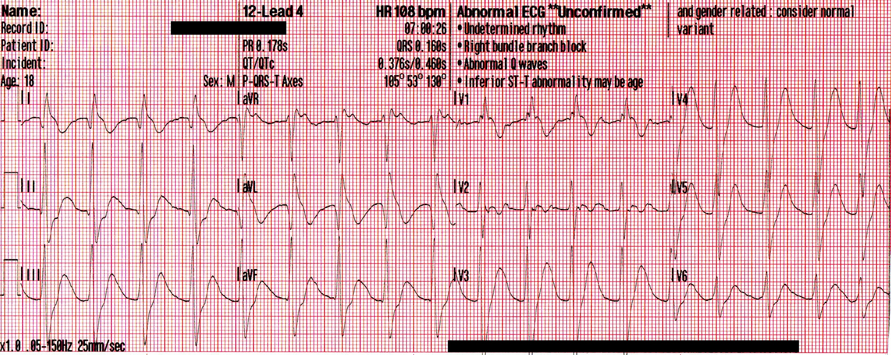
The following 12-lead was recorded following the administration of one amp of 50mEq of NaHCO3 with ongoing attempts at hyperventilation.

An irregularly-irregular wide-complex rhythm, with an apparent RBBB pattern and peaked T-waves reminiscent of hyperkalemia. This is an improvement, but there are still signs of significant sodium channel blockade.

The patient’s SpO2 improved to 100%, his blood pressure remained 85/35 [55], and despite being ventilated at a rate of 30/minute his ETCO2 remained 86mmHg. Another 50mEq of NaHCO3 is administered, and the following 12-lead is acquired:

A regular, wide-complex rhythm, with a similar QRS morphology to the previous 12-lead. The QRS is gradually narrowing, but remains pathological.

Conclusion

The crew arrived at the hospital shortly after the second amp of NaHCO3 was given. The ED staff continued administering subsequent doses of NaHCO3 , a peripheral vasopressor (norepinephrine) was initiated, and he was intubated and placed on a ventilator. Initial arterial blood gases revealed a pH of <6.8, pCO2 of >100mmHg, and a lactate of >20mmol/L. He was sent for a CT-head, which revealed no obvious findings of hemorrhage or anoxic brain injury.

He was admitted to ICU, and his repeat ABG thirty minutes later revealed an improved pH of 7.28 and pCO2 of 48mmHg. Unfortunately, no further follow-up was made available to the author.

Discussion

The critically ill toxicology patient can present many unique challenges to prehospital and ED professionals alike. Obstacles often present themselves simultaneously, including airway compromise, cardiac dysrhythmias, and hemodynamic collapse. In an unconscious patient, this is further complicated by unknown co-ingestions, quantities, and comorbidities.

This patient’s presentation can likely be explained by the complex interaction between each of the substances that were ingested. Cocaine mixed with alcohol forms cocaethylene when metabolized by the liver; a substance that’s significantly more cardiotoxic, and possesses a half-life 3-5 times that of cocaine alone. Amongst it’s multiple mechanisms, it acts as a Class Ic sodium-channel blocker, which is represented on the ECG as a progressive widening of the QRS complexes, and the development of an extreme rightward axis in the frontal plane. These channel-toxic effects are amplified by increases in heart rate and decreases in pH – two elements that are found in spades for this young man.

The deleterious effects of the cocaethylene, combined with the ingestion of significant amounts of dextromethorphan; an antitussive and a NMDA-receptor antagonist;  would likely result in euphoria, tachycardia, hypertension, dissociation, a decreasing level of consciousness, and potentially severe serotonin syndrome. Hyperthermia, tachycardia, and a disrupted respiratory drive would lead to hypercapnia, worsening acidosis, and a decreased seizure threshold. Left unchecked, this would predictably spiral into a self-perpetuating loop, inevitably resulting in profound shock and hemodynamic collapse.

Treating a patient like this with intravenous sodium bicarbonate (NaHCO3) provides a multi-pronged attack. Following administration, there’s an rapid dissociation of NaHCO3 into Na + HCO3. The extra sodium acts to “overload” the sodium-channels blocked, while the bicarbonate acts as a buffer and binds with free hydrogen (H+) ions to form Carbonic Acid (H2CO3), which then dissociates into water and carbon dioxide, expressed as HCO3 + H H2CO3 H2O + CO2. This allows for respiratory correction of the acidosis, and the subsequent alkalinization of the blood helps to reduce the channel-toxic effects of the cocaine.  It should be noted, however, that this requires an increased rate of ventilation to ensure adequate elimination of the rising CO2 levels that will follow.

In a case as advanced as this one, where severe decompensated shock has developed, stabilization becomes a delicate and complex hurdle. Since our initial treatments are aimed at alkalinization of the blood to reduce cardiotoxicity, there is a resultant left-shift of the oxyhemoglobin dissociation curve, and that leads to a decreased ability for oxygen to offload from the hemoglobin at the level of the tissue beds. This could potentially hamper our attempts to correct the massive hypoxia that’s developed, and so management is usually targeted at a pH of no higher than 7.50-7.55.

Intubation of this patient would also prove delicate, since critical hypotension and acidosis would likely be worsened by the use of most induction agents or paralytics, forcing providers to classify this as a physiologically difficult airway. For this reason, airway management should likely be accomplished using a resuscitate-before-you-intubate approach. Fluid resuscitation should be well underway before RSI, push-dose pressors should be at the ready, and providers should be aware that there’s a high-likelihood of this patient requiring vasopressor support, despite receiving a 20ml/kg crystalloid bolus.

In conclusion, the critically ill mixed-overdose patient requires aggressive yet calculated emergency management from first responders and physicians alike. A clinical understanding of the pathophysiology, as well as the implications of each aspect of treatment, is vitally important in caring for each of these patients.

Further reading on the subject

Cocaine Overdose Presents with Wide Complex TachycardiaAlec Weir, M.D. ACLSMedicalTraining.com/Blog (2016)

Role of voltage-gated sodium, potassium and calcium channels in the development of cocaine-associated cardiac arrhythmiasMichael E. O’Leary & Jules C. Hancox. British Journal of Clinical Pharmacology (Oct 2009)

Current Concepts: The Serotonin SyndromeEdward W. Boyer M.D., Ph.D., Michael Shannon M.D., M.P.H. NEJM (2005)

Treatment of patients with cocaine-induced arrhythmias: bringing the bench to the bedsideRobert S Hoffman Br J Clin Pharmacol. (2010)

Tricyclic Overdose (Sodium-Channel Blocker Toxicity) – Edward Burns, M.D. LifeInTheFastLane.com

CPR First? Or Defibrillation First?

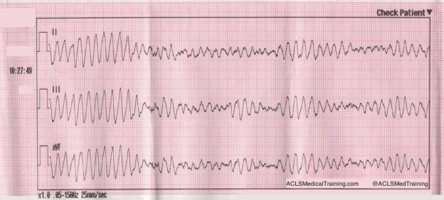
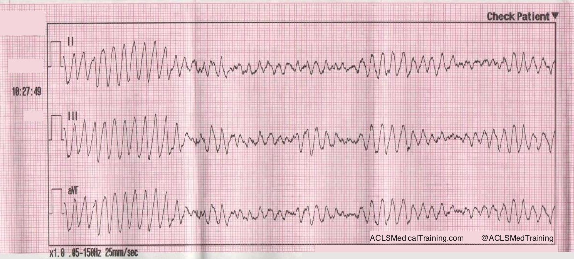
Ventricular Fibrillation is considered the most favorable cardiac arrest rhythm, and if treated promptly can result in ROSC with a favorable neurological outcome. Most survival rates are reported using witnessed arrest with a shockable rhythm as opposed to asystole or PEA, as the outcomes of these rhythms are comparatively very poor.

The Resuscitation Academy mantra “everyone in VF survives” has been adopted by many EMS systems around the world to emphasize that these patients can and do survive, and it’s up to us to save them.

Major advances have been made over the past 10 years but CPR and defibrillation are still the bedrock of resuscitation science. The attributes of high-quality CPR were re-affirmed in the 2015 AHA ECC Guidelines.

  • Ensuring adequate rate (100-120)
  • Ensuring adequate depth (2 to 2.4” or 5 to 6 cm)
  • Allowing full chest recoil (avoid leaning)
  • Minimizing interruptions to chest compressions
  • Avoiding excessive ventilations

Is CPR Before Defibrillation Dogmatic?

In the context of a witnessed arrest by a trained first responder or bystander who has an AED or manual defibrillator, the importance of early defibrillation is irrefutable. We have been told repeatedly that early defibrillation saves lives.

I initially began my research under the assumption that providing 1.5 to 3 minutes of CPR before defibrillation provides oxygen and nutrients to the heart therefore making defibrillation more likely to be successful. However, recent evidence suggests that performing chest compressions while setting up the defibrillator and charging the capacitor may be adequate.

A “CPR first” approach is rooted in evidence suggesting the existence of 3 time-sensitive phases of VF arrest.

  1. Electrical phase (0-4 minutes)
  2. Circulatory phase (5-10 minutes)
  3. Metabolic Phase (> 10 minutes)

Researchers suggested that a period of CPR prior to defibrillation might confer a benefit during the so-called “circulatory phase” of the cardiac arrest.

Evolution of American Heart Association Recommendations

Because it is rare for EMS to arrive on scene during the electrical phase, the 2005 AHA ECC Guidelines made this recommendation:

When an out-of-hospital cardiac arrest is not witnessed by EMS personnel, they may give about 5 cycles of CPR before checking the ECG rhythm and attempting defibrillation (Class IIb). One cycle of CPR consists of 30 compressions and 2 breaths. When compressions are delivered at a rate of about 100 per minute, 5 cycles of CPR should take roughly 2 minutes (range: about 1½ to 3 minutes). This recommendation regarding CPR prior to attempted defibrillation is supported by 2 clinical studies (LOE 2, LOE 3) of adult out-of-hospital VF SCA. In those studies when EMS call-to-arrival intervals were 4 to 5 minutes or longer, victims who received 1½ to 3 minutes of CPR before defibrillation showed an increased rate of initial resuscitation, survival to hospital discharge, and 1-year survival when compared with those who received immediate defibrillation for VF SCA. One randomized study, however, found no benefit to CPR before defibrillation for non-paramedic-witnessed SCA.

Fast forward 10 years to the 2015 Guidelines.

Observational clinical studies and mechanistic studies in animal models suggest that CPR under conditions of prolonged untreated VF might help restore metabolic conditions of the heart favorable to defibrillation…others have suggested that prolonged VF is energetically detrimental to the ischemic heart, justifying rapid defibrillation attempts regardless of the duration of arrest.

Evidence summary

Five RCTs, 4 observational cohort studies, 3 meta-analyses, and 1 subgroup analysis of an RCT addressed the question of CPR before defibrillation. The duration of CPR before defibrillation ranged from 90 to 180 seconds, with the control group having a shorter CPR interval lasting only as long as the time required for defibrillator deployment, pad placement, initial rhythm analysis, and AED charging. These studies showed that outcomes were not different when CPR was provided for a period of up to 180 seconds before attempted defibrillation compared with rhythm analysis and attempted defibrillation first for the various outcomes examined, ranging from 1-year survival with favorable neurologic outcome to ROSC. Subgroup analysis suggested potential benefit from CPR before defibrillation in patients with prolonged EMS response intervals (4 to 5 minutes or longer) and in EMS agencies with high baseline survival to hospital discharge, but these findings conflict with other subset analyses.  Accordingly, the current evidence suggests that for unmonitored patients with cardiac arrest outside of the hospital and an initial rhythm of VF or pVT, there is no benefit from a period of CPR of 90 to 180 seconds before attempted defibrillation.

Specifically, the ROC PRIMED trial concluded that:

Among patients who had an out-of-hospital cardiac arrest, we found no difference in the outcomes with a brief period, as compared with a longer period, of EMS-administered CPR before the first analysis of cardiac rhythm.

The ROC Investigators subsequently found that EMS systems with a VF survival rate < 20% appeared to do better with an “analyze first” strategy. Conversely, EMS systems with a VF survival rate > 20% appeared to do better with a “analyze late” strategy.

Can the VF Waveform Determine the Likelihood of Successful Defibrillation?

Ventricular fibrillation sometimes begins as ventricular tachycardia, and if left untreated deteriorates into fine VF. Fine VF predictably results in conversion to asystole or continued VF, but rarely to a perfusing rhythm.

Berg et al. performed a randomized, controlled trial using animals. After inducing VF in swine for 8 minutes, they were randomly assigned to either immediate defibrillation, or defibrillation provided after 90 seconds of CPR. Nine out of 15 attained ROSC in the CPR first group, and zero out of 15 who were defibrillated first resulted in ROSC. Their conclusion?

Pre-countershock CPR can result in substantial physiologic benefits and superior response to initial defibrillation attempts compared with immediate defibrillation in the setting of prolonged ventricular fibrillation.

Additionally, they determined there was a mathematical relationship between the VF waveform and chances of successful defibrillation. The animals who received CPR first had a much higher median frequency, and a much higher rate of ROSC than those that did not.

In the field, whether or not VF is “fine” or “coarse” is typically based on visual inspection of the waveform. What if there was a way to accurately determine which patients would benefit from defibrillation and those that would not, thus eliminating unnecessary pauses and ineffective shocks?

Callaway et al. and Eftestol et al. supported the theory that VF frequency and amplitude could be used to determine which patients will respond to countershock.

Eftestol et al. concluded:

CPR done by professionals can improve the chance for ROSC and ultimate survival of patients with prolonged cardiac arrest and significantly deteriorated myocardium. This study also indicates that CPR periods of 3 minutes might be better for the myocardium than shorter periods. Finally, together with the studies showing rapid deterioration of the myocardium in even a few seconds without CPR after a cardiac arrest, it gives the important message that periods without CPR (for ECG analysis, defibrillation charging, pulse checks, intubation attempts, etc) should be kept to a minimum. This is frequently not the case clinically.

As promising as this may have seemed, an article in Circulation by Freese et al. evaluated the theory of defibrillation based on waveform analysis, and the results were disappointing.

Use of a waveform analysis algorithm to guide the initial treatment of out-of-hospital cardiac arrest patients presenting in VF did not improve overall survival compared with a standard shock-first protocol. Further study is recommended to examine the role of waveform analysis for the guided management of VF.

The Bottom Line

The totality of the evidence suggests that defibrillation as soon as practicable (with the caveat that high quality chest compressions are performed while setting up the defibrillator) is equivalent to a prescribed interval of CPR prior to the first shock in most instances.

EMS systems that measure the “patient’s side to first shock” interval know that it usually takes at least 1 minute to power on the defibrillator, extend the cables, attach the pads, charge the capacitor, and deliver the shock. During that interval, there’s no reason that the patient can’t receive continuous chest compressions.

One benefit to emphasizing a “shock as soon as possible” approach is that it’s the same for EMS-witnessed cardiac arrest.

Alternatively, defibrillation can be delivered after the first 2-minute cycle. It seems likely that CPR quality plays a more important role than the exact timing of the first shock.

References

Baker PW, Conway J, Cotton C, Ashby DT, Smyth J, Woodman RJ, Grantham H; Clinical Investigators. Defibrillation or cardiopulmonary resuscitation first for patients with out-of-hospital cardiac arrests found by paramedics to be in ventricular fibrillation? A randomised control trial. Resuscitation. 2008;79:424–431. doi: 10.1016/j.resuscitation.2008.07.017.

Bradley SM, Gabriel EE, Aufderheide TP, Barnes R, Christenson J, Davis DP, Stiell IG, Nichol G; Resuscitation Outcomes Consortium Investigators. Survival increases with CPR by Emergency Medical Services before defibrillation of out-of-hospital ventricular fibrillation or ventricular tachycardia: observations from the Resuscitation Outcomes Consortium. Resuscitation. 2010;81:155–162. doi: 10.1016/j. resuscitation.2009.10.026.

Cobb LA, Fahrenbruch CE, Walsh TR, Copass MK, Olsufka M, Breskin M, Hallstrom AP. Influence of cardiopulmonary resuscitation prior to defibrillation in patients with out-of-hospital ventricular fibrillation. JAMA. 1999;281:1182–1188.

Freese J, Jorgenson D, Liu P et al. Waveform Analysis-Guided Treatment Versus a Standard Shock-First Protocol for the Treatment of Out-of-Hospital Cardiac Arrest Presenting in Ventricular Fibrillation: Results of an International Randomized, Controlled Trial. Circulation. 2013;128(9):995-1002. doi:10.1161/circulationaha.113.003273.

Gilmore C, Rea T, Becker L, Eisenberg M. Three-Phase Model of Cardiac Arrest: Time-Dependent Benefit of Bystander Cardiopulmonary Resuscitation. The American Journal of Cardiology. 2006;98(4):497-499. doi:10.1016/j.amjcard.2006.02.055.

Hayakawa M, Gando S, Okamoto H, Asai Y, Uegaki S, Makise H. Shortening of cardiopulmonary resuscitation time before the defibrilla- tion worsens the outcome in out-of-hospital VF patients. Am J Emerg Med. 2009;27:470–474. doi: 10.1016/j.ajem.2008.

Huang Y, He Q, Yang LJ, Liu GJ, Jones A. Cardiopulmonary resuscitation (CPR) plus delayed defibrillation versus immediate defibrillation for out-of-hospital cardiac arrest. Cochrane Database Syst Rev. 2014;9:CD009803. doi: 10.1002/14651858.CD009803.pub2.

Jacobs IG, Finn JC, Oxer HF, Jelinek GA. CPR before defibrillation in out-of-hospital cardiac arrest: a randomized trial. Emerg Med Australas. 2005;17:39–45. doi: 10.1111/j.1742-6723.2005.00694.x.

Kleinman M, Brennan E, Goldberger Z et al. Part 5: Adult Basic Life Support and Cardiopulmonary Resuscitation Quality. Circulation. 2015;132(18 suppl 2):S414-S435. doi:10.1161/cir.0000000000000259.

Koike S, Tanabe S, Ogawa T, Akahane M, Yasunaga H, Horiguchi H, Matsumoto S, Imamura T. Immediate defibrillation or defibrillation after cardiopulmonary resuscitation. Prehosp Emerg Care. 2011;15:393–400. doi: 10.3109/10903127.2011.569848.

Ma MH, Chiang WC, Ko PC, Yang CW, Wang HC, Chen SY, Chang WT, Huang CH, Chou HC, Lai MS, Chien KL, Lee BC, Hwang CH, Wang YC, Hsiung GH, Hsiao YW, Chang AM, Chen WJ, Chen SC. A randomized trial of compression first or analyze first strategies in patients with out-of-hospital cardiac arrest: results from an Asian community. Resuscitation. 2012;83:806–812. doi: 10.1016/j.resuscitation.2012.01.009.

Meier P, Baker P, Jost D, Jacobs I, Henzi B, Knapp G, Sasson C. Chest compressions before defibrillation for out-of-hospital cardiac arrest: a meta-analysis of randomized controlled clinical trials. BMC Med. 2010;8:52. doi: 10.1186/1741-7015-8-52.

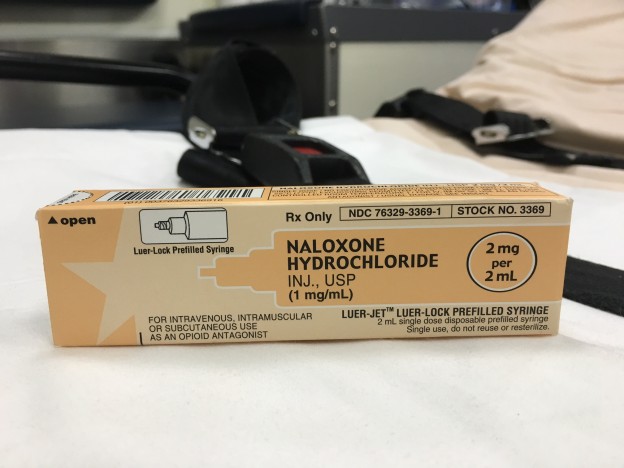
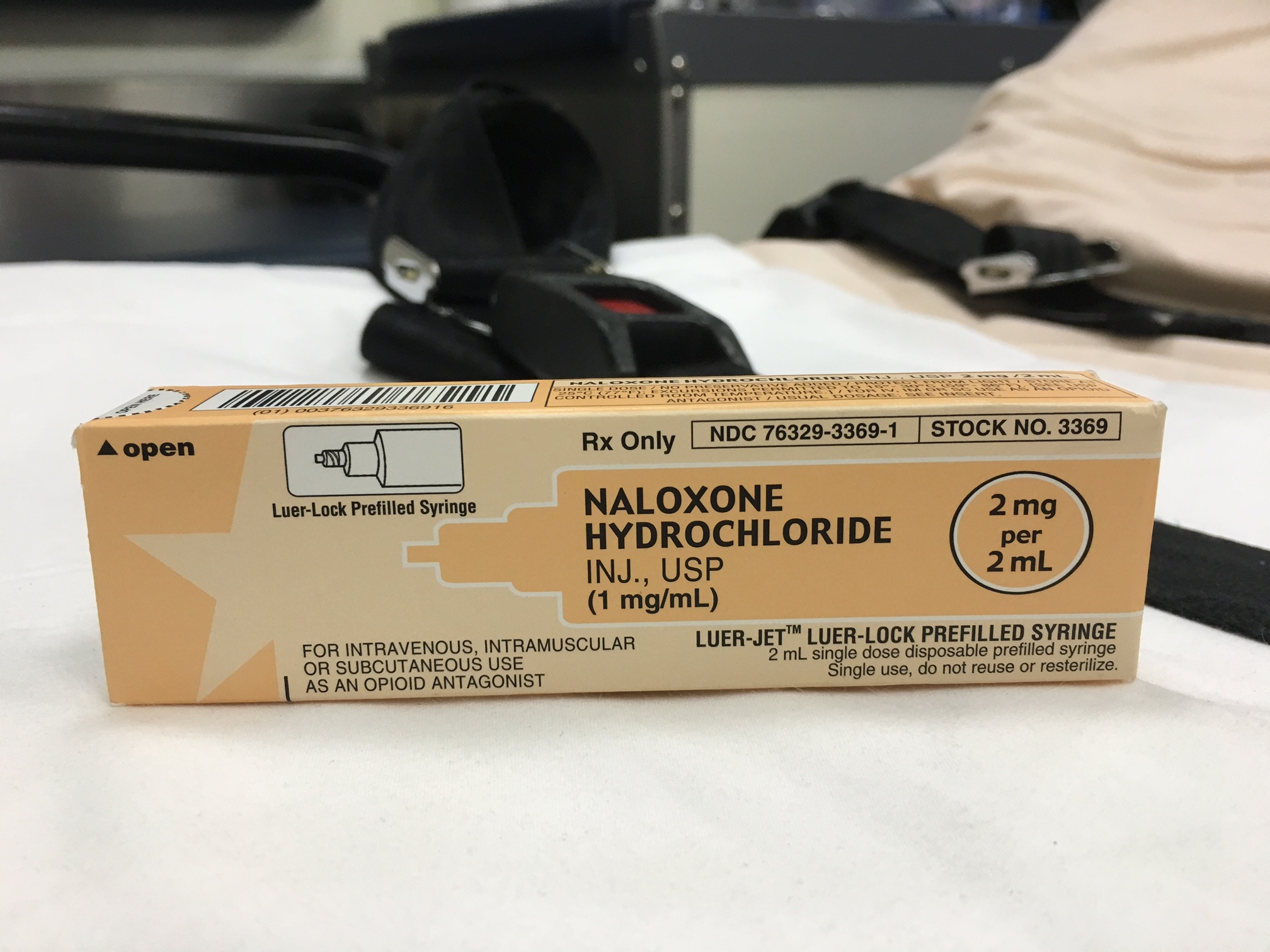
Naloxone

Is there an Irrational Fear of Naloxone?

Naloxone

In the U.S. there has been a 286% increase in heroin-related overdose deaths since 2002. In 2014, a total of 47,055 drug overdose deaths occurred. Our EMS systems and emergency departments have felt the surge, along with a corresponding increase in the use of the drug naloxone.

Community officials have taken the initiative to reduce the number of overdose deaths by making naloxone available through non-traditional means. However, this has not been particularly well received by members of the medical community, particularly pre-hospital providers.

Discussion of the proper use of naloxone often sparks a passionate, sometimes contentious debate, usually focused around the deleterious side effects and inappropriate use of naloxone.

Hopefully, a review of the literature will make providers less apprehensive about the expanded use of naloxone and spark a healthy debate of the current standard of care.

Treatment of an opioid overdose

A common strategy for treating an opioid-related overdose is to initiate BVM ventilation to treat respiratory depression and then titrating naloxone until the patient’s respiratory effort becomes adequate but not to the point of the patient becoming lucid.

Should patients be given enough naloxone to regain consciousness?

There was a time when I passionately advocated for allowing patients to remain somnolent in the setting of a suspected opioid overdose, but I changed my mind after encountering this emergency more frequently.

Like many things in medicine, it sounds good in theory, but it doesn’t translate easily into practice. Almost every attempt I made at titrating naloxone until the respiratory depression improved resulted in the patient becoming completely lucid.

What about adverse outcomes associated with naloxone administration?

Many emergency providers are apprehensive about giving naloxone due to undesirable side effects. Patients regaining consciousness after naloxone administration may experience many symptoms that are collectively referred to as Acute Withdrawal Syndrome (AWS).

Patients with AWS may exhibit nausea, vomiting, tachycardia, diarrhea, hypertension, nervousness, and restlessness. The degree of withdrawal symptoms is relatively proportional to the amount of naloxone given, so smaller amounts will normally result in less serious withdrawal symptoms.

Rare but serious complications like cardiac arrest, seizures, acute pulmonary edema, and violent behavior are sometimes offered as reasons for not using naloxone (or not giving enough for the patient to regain consciousness). However, it is likely that these complications have been overstated, as the evidence has not been reproducible.

Osterwalder (1996) investigated subjects treated with naloxone from 1991-1993. Six out of 453 patients experienced severe adverse effects. One suffered asystole, three generalized convulsions, one pulmonary edema, and one violent behavior.

However, Burris (2000) reported in the International Journal of Drug Policy that “more recent research suggests that complications are exceedingly rare, that past reports of complications may have been erroneous, or that complications occur, if at all, in patients with pre-existing heart disease”.

Yearly et al. (1990) conducted a retrospective study of over 800 prehospital records of patients who received initial IV doses of 0.4-0.8 mg of naloxone and found that no patients experienced ventricular tachycardia, fibrillation, or asystole. There was one generalized tonic-clonic seizure in a patient with a history of seizures. The authors concluded that smaller doses of naloxone are not warranted.

What if a patient refuses transport after regaining consciousness?

Typically patients are transported to the hospital after regaining consciousness following the administration of naloxone. However some patients refuse, against the advice of treating paramedics.

One might expect there to be a high mortality for patients who refuse transport, since it’s widely known that the half-life of naloxone is shorter than the half-life of the opioid. However, current data suggest that a patient can refuse transport without serious consequences.

In San Antonio, Wampler et al. (2011) conducted a review of 595 patients treated with naloxone in a large fire-based EMS system who refused transport to the hospital. The San Antonio protocol consisted of giving naloxone, 2 mg IM, 2 mg IV, and an additional 2mg IM with patient consent. None were found in the Medical Examiner’s Office database two-days after refusing transport. Although 9 of the patients subsequently died, the shortest time interval was four days after treatment.

Vike et al (2003) conducted another retrospective review comparing prehospital and medical examiner databases. In the prehospital database there were 998 patients who had received naloxone and refused transport. In the medical examiner’s database there were 601 recorded opioid overdose deaths. None of them had been treated with naloxone within 12 hours of death.

Conclusion

  • Naloxone is a safe and effective treatment for opioid overdose.
  • Expanded use of naloxone is unlikely to cause an increase in adverse outcomes.
  • Criteria should be established to help predict patients that can safely refuse transport to the hospital.

References

Burris S, Norland J, Edlin B. Legal aspects of providing naloxone to heroin users in the United States. International Journal of Drug Policy. 2001;12(3):237-248.

Kim D, Irwin K, Khoshnood K. Expanded Access to Naloxone: Options for Critical Response to the Epidemic of Opioid Overdose Mortality. Am J Public Health. 2009;99(3):402-407.

Opioid OD patients revived with naloxone who refuse further treatment do not die. EMSWorld.com. 2016. Available at: http://www.emsworld.com/article/10284002/opioid-od-patients-revived-with-naloxone-who-refuse-further-treatment-do-not-die. Accessed July 12, 2016.

Wermeling D. Review of naloxone safety for opioid overdose: practical considerations for new technology and expanded public access. Therapeutic Advances in Drug Safety. 2015;6(1):20-31.

Wampler D, Molina D, McManus J, Laws P, Manifold C. No Deaths Associated with Patient Refusal of Transport After Naloxone-Reversed Opioid Overdose. Prehospital Emergency Care. 2011;15(3):320-324.

Boyer E. Management of Opioid Analgesic Overdose. New England Journal of Medicine. 2012;367(2):146-155.

Vilke G, Buchanan J, Dunford J, Chan T. Are heroin overdose deaths related to patient release after prehospital treatment with naloxone?. Prehospital Emergency Care. 1999;3(3):183-186.

SVT with Aberrancy or Ventricular Tachycardia?

What is SVT with aberrancy?

The term “SVT with aberrancy” tends to throw many providers off so let’s start by defining SVT using the 2015 ACC/AHA/HRS Guidelines as reference.

“An umbrella term used to describe tachycardias (atrial and/or ventricular rates in excess of 100 bpm at rest), the mechanism of which involves tissue from the His bundle or above. These SVTs include inappropriate sinus tachycardia, AT (including focal and multifocal AT), macroreentrant AT (including typical atrial flutter), junctional tachycardia, AVNRT, and various forms of accessory pathway-mediated reentrant tachycardias. In this guideline, the term does not include AF.”

This is important because many of us were taught a narrow complex rhythm “must be SVT if the rate is over 150,” which can lead to inappropriate therapies. In reality, sinus tachycardia is a form of SVT, and the rate can easily exceed 150. A good rule of thumb to estimate the maximum sinus rate is 220 minus age but that can vary by 10-15%, which is a lot.

What most people really mean when they call a rhythm “SVT” is AV Nodal Reentrant Tachycardia or AVNRT, which is a reentrant rhythm in or around the AV node. This arrhythmia is usually stable and the prognosis is much more favorable than VT. It is usually treated with vagal maneuvers or adenosine.

What does aberrancy mean?

You can think of “aberrancy” as abnormal conduction. When something is aberrant it “departs from the right, normal, or usual course.”

Because the right bundle branch tends to have a slightly longer refractory period than the left bundle branch, at higher rates the right bundle branch may not be fully recovered from the previous cardiac cycle, which results in a right bundle branch block pattern.

Even though right bundle branch block aberrancy is more common than left bundle branch block aberrancy, both are possible. Additionally, we know that many patients have underlying bundle branch block, including bifascicular block, at baseline.

When a patient with a bundle branch block experiences SVT the result is a wide complex tachycardia.

Can you differentiate between SVT with aberrant conduction and VT?

The short answer is yes, but it can be very difficult, and even experienced clinicians can misdiagnose VT as SVT with aberrancy!

This can lead to clinical misadventure. In particular, treating a wide complex tachycardia with a calcium channel blocker is a dangerous decision that could have fatal consequences for your patient.

There are good criteria to help rule-in, or tip the scales in favor of VT, but none to safely rule-out VT.

See also: Myths and Cognitive Biases in Interpretation of  Wide Complex Tachycardias

Consider the following case

EMS is dispatched to an 83-year-old female who contacts 9-1-1 after she wakes up with a “racing heart” and shortness of breath.

Past medical history includes myocardial infarction and hypertension.

On initial assessment the patient is found to be alert and oriented to person, place, time, and event. The skin is pale but warm and dry. Radial pulses are very rapid but surprisingly strong. Breath sounds are clear bilaterally.

She is placed on the cardiac monitor and the following rhythm strip is obtained.

Figure 1: There is a wide and regular complex tachycardia at a rate of ~ 230 bpm.

The patient is placed on oxygen via nasal cannula and IV access is established while vital signs are obtained.

  • RR: 24
  • HR: Too fast to count
  • NIBP: 112/72
  • SpO2: 97%
  • Temp: 98.3 F / 36.8 C

Why should you presume that this rhythm is ventricular tachycardia?

  • VT accounts for 80% of all cases of WCT
  • If the patient has a previous cardiac history, the predictive value can go up over 90%
  • An age greater than 35 years has a sensitivity of 92%

Treatment

A 12 lead ECG is obtained.

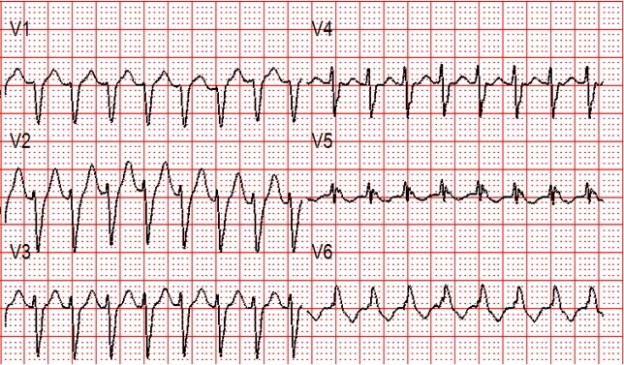
Figure 2: There is a regular wide complex tachycardia at a rate of about 230 without sinus P waves. There is a LBBB pattern in lead V1. However, we would not consider this to be a “typical” LBBB pattern due to the normal axis in the frontal plane and the presence of a small S-wave in lead I.

Amiodarone 150 mg is given over 10 minutes.

A rhythm change is noted and the following 12-lead ECG is obtained.

Figure 3: Now there is sinus tachycardia with virtually identical QRS morphology.

Once the patient converts to sinus tachycardia (and after a sigh of relief) paramedics compare the two 12-lead ECGs. The axis and QRS morphology are noted to be exactly the same.

The diagnosis? SVT with aberrancy!

It is safe to conclude that this patient had a conduction defect at baseline, which is what caused the complexes to be wide during the tachycardia.

Retrospectively, adenosine would have been safe and likely effective. In many cases it can be considered as a first line therapy for undifferentiated wide complex tachycardia, and may have some diagnostic utility when considered in the context of other findings.

Summary

  • Wide complex tachycardias should be presumed to be VT until proven otherwise
  • Obtain a 12-lead ECG before and after treatment to help aid in the diagnosis
  • Unstable WCT requires immediate synchronized cardioversion (when the symptoms are believed to be due to the heart rhythm)
  • Consider adenosine as an initial therapy for an undifferentiated wide complex tachycardia

References

Alzand BCrijns H. Diagnostic criteria of broad QRS complex tachycardia: decades of evolution. Europace. 2010;13(4):465-472

Neumar R, Otto C, Link M et al. Part 8: Adult Advanced Cardiovascular Life Support: 2010 American Heart Association Guidelines for Cardiopulmonary Resuscitation and Emergency Cardiovascular Care. Circulation. 2010;122(18_suppl_3):S729-S767