SVT with Aberrancy or Ventricular Tachycardia?
Ask a non-cardiologist how to tell SVT with aberrancy from ventricular tachycardia (VT) and you may be met with a blank stare. Distinguishing between wide complex tachycardias (WCT) such as SVT with aberrancy and VT can be a challenging task, even for experienced healthcare providers, which is why rhythm recognition is a central focus of ACLS certification training.
WCTs are some of the most dangerous ECG findings since VT makes up the vast majority and poses severe risks of hemodynamic decompensation. The dangers, thus, of misdiagnosing VT as SVT with aberrancy carry significant consequences, as medications used for the latter can be deadly for the former.
In this blog, we will aim to clarify the distinction between these two cardiac arrhythmias, understanding that in a given moment of new-onset VT or SVT with aberrancy, perfect certainty in rhythm interpretation is a myth.
Table of Contents
- What Is Wide Complex Tachycardia?
- What Is SVT With Aberrancy?
- What Does Aberrant Conduction Mean?
- What Is Ventricular Tachycardia?
- Why Misdiagnosing VT is Dangerous
- SVT With Aberrancy vs. VT: Key Differences to Evaluate
- Clinical Rules and Red Flags That Favor VT
- Case Example: When SVT with Aberrancy Mimics VT
- Treatment Approach for Undifferentiated Wide Complex Tachycardia
- Practical Takeaways for Clinicians
- Take the Next Step in Cardiac Care With ACLS Medical Training
What Is Wide Complex Tachycardia?
Wide complex tachycardia is an accelerated heart rate (>100 bpm) with a widened QRS (>120 milliseconds). For a step-by-step breakdown of treatment pathways for wide complex tachycardia, including antiarrhythmics and synchronized cardioversion, see our guide to wide complex tachycardia treated with amiodarone and synchronized cardioversion .
Our difficulty lies in that both SVT with aberrancy and VT match this description on an ECG tracing. Since this is the case, clinicians must err on the side of the more dangerous rhythm: VT. The rule is always to “treat WCT as VT until proven otherwise.”
A more detailed discussion of medication and cardioversion strategies for wide complex tachycardia can be found in our guide on wide complex tachycardia treated with amiodarone and synchronized cardioversion .
What Is SVT With Aberrancy?
It’s not uncommon for clinicians to answer the question “Is wide complex tachycardia always VT?” with a yes, but this is a mistake. An SVT with aberrancy ECG interpretation can easily be confused as VT because they share a broadly similar morphology with accelerated rates, widened QRS complexes, and little-to-no visualization of P waves.
While traditional SVT commonly presents with a narrow QRS, SVT with aberrancy is distinguished as a tachyarrhythmia in which impulses originating above the ventricles result in a wide QRS morphology. This is due to impaired intraventricular conduction and occurs via two primary mechanisms: baseline conduction defects with bundle branch block (where ventricular depolarization is delayed due to having to travel via the myocardium instead of the “lightning-fast” His-Purkinje system), or rate-related aberrancy (where the SVT cycle length is shorter than the effective refractory period of a bundle branch, again producing slower depolarization through the transseptal myocardium).
What Does Aberrant Conduction Mean?
When something is aberrant, it “departs from the right, normal, or usual course.” In the context of SVT, aberrancy refers to the abnormal intraventricular conduction of a supraventricular impulse due to deviation from the normal course of conduction through the His-Purkinje system which causes a wider QRS on the ECG tracing.
Both defects cause the QRS to widen due to extended time of depolarization through the myocardium.
While aberrant conduction itself is not inherently dangerous, the potential for misdiagnosis is a significant concern. Mistaking SVT with aberrancy for VT can lead to the administration of antiarrhythmic medications or cardioversion, which can be harmful if the underlying rhythm is SVT.
What Is Ventricular Tachycardia?
The American Heart Association defines VT as a “cardiac arrhythmia of ≥3 consecutive complexes originating in the ventricles at a rate >100bpm”. Sustained VT lasts >30 seconds while nonsustained VT lasts ≥3 beats but less than 30 seconds. VT is life-threatening because it can cause hemodynamic collapse with sudden cardiac death upon initiation or at any point while the rhythm is ongoing.
Common causes of VT include:
- Ischemic heart disease
- Prior MI
- Dilated cardiomyopathy
- Structural heart disease
- Electrolyte imbalances
Because these etiologies are so common, VT accounts for the majority of wide complex tachycardias.
In terms of prognosis, VT is much more deadly than SVT which typically only causes palpitations or lightheadedness.
Why Misdiagnosing VT is Dangerous
Given the difference in mortality and treatment between VT and SVT, extreme caution must be given so that VT is not treated as SVT. In particular, antiarrhythmics and calcium channel blockers which are given to patients with SVT can be fatal if given to a patient with VT. This is because these medications can cause significant hypotension, stamping out any ongoing compensatory effort to maintain hemodynamic stability for a patient experiencing VT. Thus, it is crucial to select the safest treatment strategy for the patient and err on the side of caution for treatment modalities when the exact type of WCT is unknown.
SVT With Aberrancy vs. VT: Key Differences to Evaluate
Differentiating between SVT with aberrancy and VT can help determine the appropriate treatment. Several factors can help distinguish between the two, including the patient’s history and age: for instance, a young patient (<35 years) with a history of bundle branch block or a wide QRS complex at baseline is more of a candidate for SVT with aberrancy; in contrast, a patient >35 years with a history of heart disease, MI, or congestive heart failure years old will strongly suggest VT (85-95% predictive value).
Both SVT with aberrancy and VT are strikingly similar in appearance on a 12-lead ECG as they are both regular and rapid rhythms with similar heart rate ranges (120 to 220 [VT] vs. 150 to 250+ [SVT]). However, the ECG can still provide clues in the QRS morphology and axis findings on a 12 lead ECG to distinguish between the two: an extreme right axis deviation likely determines VT, and when all six precordial leads (V1 through V6) are monophasic with the same polarity (also known as “concordance”), VT is very likely.
Importantly, while there are diagnostic criteria to help rule in VT, there are currently no definitive criteria to safely rule out VT.
Clinical Rules and Red Flags That Favor VT
Strictly from an odds standpoint, VT accounts for about 80% of WCT while SVT only comprises about 15 to 20%. The predictive odds for VT also increase when the patient is >35 years old or has a history of MI or cardiomyopathy. Additionally, visible but inconsistent P waves in a WCT demonstrate AV dissociation (atria and ventricles firing independently) which is diagnostic of VT.
Case Example: When SVT with Aberrancy Mimics VT
Consider the following case:
EMS is dispatched to an 83-year-old female who contacts 9-1-1 after she wakes up with a “racing heart” and shortness of breath.
Past medical history includes myocardial infarction and hypertension.
On initial assessment the patient is found to be alert and oriented to person, place, time, and event. The skin is pale but warm and dry. Radial pulses are very rapid but surprisingly strong. Breath sounds are clear bilaterally.
She is placed on the cardiac monitor, and the following rhythm strip is obtained.

Figure 1 : There is a wide and regular complex tachycardia at a rate of ~ 230 bpm.
The patient is placed on oxygen via nasal cannula, and IV access is established while vital signs are obtained.
- RR: 24
- HR: Too fast to count
- NIBP: 112/72
- SpO2: 97%
- Temp: 98.3 F / 36.8 C
Why should you presume that this rhythm is ventricular tachycardia?
- VT accounts for 80% of all cases of WCT
- An age greater than 35 years has a sensitivity of >85%
- If the patient has a previous MI, the predictive value can exceed 90%
Treatment
A 12 lead ECG is obtained.
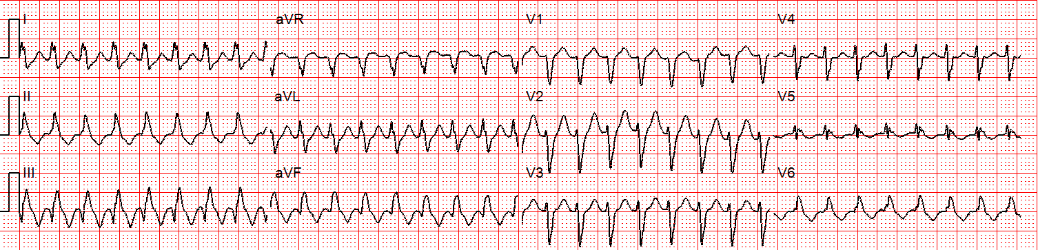
Figure 2 : There is a regular wide complex tachycardia at a rate of about 230 without sinus P waves. There is a LBBB pattern in lead V1. However, we would not consider this to be a “typical” LBBB pattern due to the normal axis in the frontal plane and the presence of a small S-wave in lead I.
Amiodarone 150 mg is given over 10 minutes.
A rhythm change is noted, and the following 12-lead ECG is obtained.
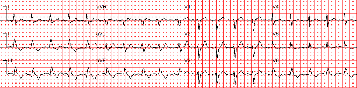
Figure 3 : Now there is sinus tachycardia with still with a virtually identical QRS morphology.
Once the patient converts to sinus tachycardia, paramedics compare the two 12-lead ECGs. The axis and QRS morphology are noted to be exactly the same. Because of this, we can confidently say the patient had SVT with aberrancy. It is safe to conclude that this patient had a conduction defect at baseline, which is what caused the complexes to be wide during the tachycardia.
Retrospectively, adenosine would have been safe and likely effective. In many cases, it can be considered as a first line therapy for undifferentiated wide complex tachycardia, and may have some diagnostic utility when considered in the context of other findings.
Treatment Approach for Undifferentiated Wide Complex Tachycardia
Ultimately, if there is confusion as to whether a WCT is VT or SVT with aberrancy, the rhythm should be treated as VT until proven otherwise. Ventricular tachycardia versus SVT treatment should prudently err on the side of VT. This is because of the difference in risk to the patient: SVT with aberrancy is rarely lethal whereas VT is a deadly rhythm.
For an unstable patient with WCT, rhythm differentiation is not truly necessary in the moment as synchronized cardioversion is the standard treatment for any unstable tachycardia regardless of origin. If the patient is stable with a known SVT, then vagal maneuvers followed by adenosine and a calcium channel blocker are advised.
Lastly, pre- and post-treatment 12-lead ECGs should be taken to compare QRS morphology for definitive determination of SVT with aberrancy versus VT. This should also be performed to determine whether myocardial ischemia is present following the episode of WCT.
Practical Takeaways for Clinicians
To summarize, do not assume that an undifferentiated WCT is SVT based on rate alone. Always consider and treat the rhythm as VT first, recognizing the dangers of doing the reverse. Consider the patient’s medical history and age, in addition to ECG tracings, to quickly consider the odds of VT in your approach to treatment. When in doubt, remember that the risk differential between the two rhythms strongly favors treating the rhythm as VT and err on the side of choosing the safest therapy.
Take the Next Step in Cardiac Care With ACLS Medical Training
Precision in distinguishing SVT with aberrancy from ventricular tachycardia is crucial for safe and timely patient care. Misdiagnosis can have severe consequences. Having a strong foundation in rhythm recognition reduces cognitive burden and empowers clinicians to act quickly when seconds matter.
Mastering ACLS arrhythmia recognition for wide complex tachycardia requires continued study and practical review of treatment algorithms. Whether you are preparing for certification or maintaining your credentials, ongoing education helps reinforce accurate ECG interpretation and sound clinical decision-making.
We offer the following online training programs:
Strengthen your skills in advanced cardiac life support and stay prepared to manage high-risk cardiac emergencies with confidence. Visit our website to get started today.
References
Al-Khatib SM, Stevenson WG, Ackerman MJ, et al. 2017 AHA/ACC/HRS guideline for management of patients with ventricular arrhythmias and the prevention of sudden cardiac death: a report of the American College of Cardiology/American Heart Association Task Force on Clinical Practice Guidelines and the Heart Rhythm Society. J Am Coll Cardiol. 2018;72(14):e91-e220. doi:10.1016/j.jacc.2017.10.054
Baerman JM, Morady F, DiCarlo LA Jr, Billman GF. Differentiation of ventricular tachycardia from supraventricular tachycardia with aberration: value of the clinical history. Ann Emerg Med. 1987;16(1):40-43. doi:10.1016/s0196-0644(87)80283-4