Transcutaneous Pacing (TCP) With and Without Capture
Transcutaneous pacing (TCP) is an emergency treatment used to manage unstable symptomatic bradycardia, but seeing pacing spikes on the ECG doesn't necessarily mean the heart is pumping effectively. Recognizing the difference between true and false capture is a critical skill taught in ACLS training , helping providers determine whether pacing is actually restoring adequate perfusion.
This guide explains how to identify true versus false capture, troubleshoot failed pacing, and improve patient outcomes in emergency situations.
Table of Contents
- EMS Response to an Unstable Bradycardia Patient
- Initial Patient Assessment and Vital Signs
- ECG Findings – High-Degree AV Block with Escape Rhythm
- Indications for Transcutaneous Pacing
- Initiation of Transcutaneous Pacing in the Field
- Apparent Capture with Persistent Hypotension
- Additional Prehospital Interventions
- Transition to Transvenous Pacing and Hospital Course
- Why Transcutaneous Pacing Often Fails to Achieve True Capture
- What is False Capture in Transcutaneous Pacing
- Identifying True Electrical and Mechanical Capture
- Tools to Confirm Mechanical Capture
- Common Reasons for Failure to Capture
- Practical Tips for Successful Transcutaneous Pacing
- Key Clinical Takeaways for EMS and Emergency Providers
EMS Response to an Unstable Bradycardia Patient
EMS is dispatched to an apartment for a 68-year-old male who was reported unconscious by his girlfriend. Upon arrival, EMS providers find the patient lying on the kitchen floor with no response to painful stimuli. The patient’s skin is pale, cool and clammy. Occasional snoring respirations are noted, and a slow carotid pulse is present. The girlfriend states that the patient’s medical history includes heart failure, hypertension and hyperlipidemia. For the unconscious patient with these findings, it is critical to perform an urgent assessment and early intervention.
Initial Patient Assessment and Vital Signs
As always, begin with ABC’s (Airway, Breathing, Circulation). The airway is apparently clear, despite the patient snoring and being unresponsive, and vital signs are as follows:
- RR: 8
- HR: 28
- NIBP: 76/41
- SpO2: Not registering
- Temp: 96.1 F / 36.7 C
- BGL: 101 mg/dL
Severe symptomatic bradycardia with hypotension is a significant and life-threatening emergency. This patient currently has inadequate cardiac output from a low heart rate, which leads to poor perfusion pressure and a shock state that is not being compensated by the sympathetic nervous system.
ECG Findings – High-Degree AV Block with Escape Rhythm
The cardiac monitor is attached and the following rhythm is shown:
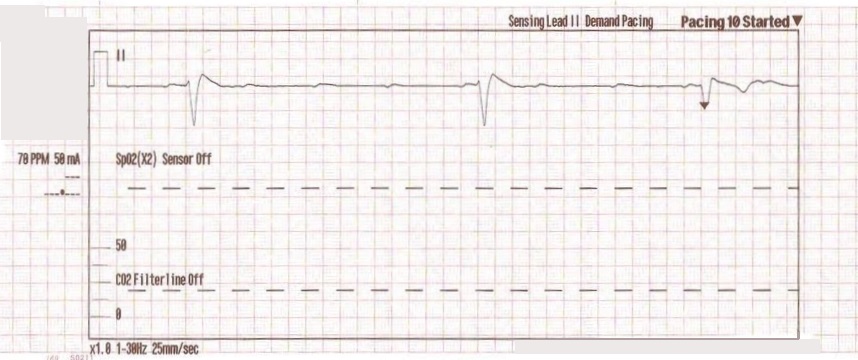
High degree AV block with wide complex escape rhythm.
Because of the immediate dangers this rhythm carries, namely hypotension and shock, plus the potential danger for transitioning to full asystole, immediate intervention with transcutaneous pacing is mandatory to prevent cardiovascular collapse and death.
Indications for Transcutaneous Pacing
TCP is indicated in unstable bradycardia where the administration of atropine is either ineffective or not indicated. In cases of High-Degree AV Block, atropine is typically not indicated since activation of the SA node will have no benefit on the blocked AV node – remember that high degree blocks are an AV node problem and no amount of SA node firing will fix that.
Thus, transcutaneous pacing is a rapid and effective alternative that is indicated for patients with hypotension in the setting of severe bradycardia. However, it is meant to be a temporary measure due to the potential for severe discomfort, skin injury, and failure to achieve consistent mechanical capture (which is discussed further below).
Initiation of Transcutaneous Pacing in the Field
Going back to our scenario, the adult pads are placed and transcutaneous pacing is initiated with the following findings on the monitor:
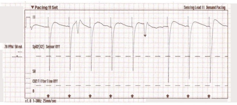
The transcutaneous pacer is set for 70 PPM at 50 mA. Pacing spikes are visible with what appear to be large, corresponding QRS complexes.
To properly apply pacing pads, place them directly on the chest, historically in the anterior-lateral positions, although anterior-posterior pad placement is now recommended since it allows for more effective capture. Chest hair may be shaven as necessary. Once pads are applied, the transcutaneous pacer machine may be turned on.
Next, the machine should be set to “demand” mode (which inhibits pacing in the presence of intrinsic cardiac beats), with the rate set anywhere from 60 to 80bpm. The pacer current should be set at the lowest rate that achieves mechanical capture, meaning that each pacer impulse results in a complete cardiac cycle on the ECG . In conscious patients, this is achieved by increasing the current by 5 or 10 mA at a time until capture is seen, but in unconscious patients, providers should start at the maximum current and titrate down until the capture threshold is found. Once the capture threshold is found, increase the current by 10 mA to ensure a safety “buffer” to ensure continued capture.
Pacing spikes appear as sharp vertical lines indicating that an electrical impulse has been delivered. In true capture, every pacing spike will be followed by a wide QRS complex which indicates that an impulse has successfully travelled across the cardiac conduction system.
Apparent Capture with Persistent Hypotension
Going back to our scenario, the patient’s blood pressure improves slightly to 84/47 and a carotid “pulse” is felt. Nonetheless, paramedics are still concerned about the patient’s hypotension. It is possible, and not infrequent, that apparent QRS complexes are present on the ECG monitor but hypoperfusion still persists.
Additionally, the patient may show false signs of improvement due to skeletal muscle contraction. For example, the high-voltage current stimulates the pectoral and intercostal muscles, resulting in jerking of the chest wall and arms which can deceptively appear as the patient recovering. It may also cause a false carotid pulse to be felt which is actually a muscle impulse from contractions in the neck.
Additional Prehospital Interventions
For the patient in our scenario, EMS providers continued pacing the patient at the above settings and performed multiple adjunctive treatments including:
- IO access obtained in right proximal tibia.
- 0.5 mg of Atropine administered x 3.
- 1L of normal saline run wide open with an additional IV line established in the left lower extremity.
These therapies are often performed simultaneously by EMS or ICU teams since each intervention is dependent on the performance of the others for reversing hypoperfusion and shock.
Transition to Transvenous Pacing and Hospital Course
While en route to the hospital, the patient begins to move and reaches for the pacing pads. However, he is still non-verbal and does not follow commands. On arrival at the hospital ED, the patient is transitioned to transvenous pacing before being transferred to the ICU. Once in ICU, the patient remains dangerously hypotensive in spite of dobutamine and norepinephrine drips. Due to the patient’s severe and prolonged shock state, the patient codes multiple times and eventually expires from multi-organ failure.
While often reversible, prolonged hypoperfusion from symptomatic bradycardia with low cardiac output can eventually lead to permanent organ damage and death.
Why Transcutaneous Pacing Often Fails to Achieve True Capture
The goal of pacing is to obtain successful capture, both electrical and mechanical. Electrical capture is achieved when a pacing impulse causes depolarization of the ventricles, demonstrated by a wide QRS followed by a T wave on the ECG monitor. Mechanical capture occurs when the myocardium contracts, demonstrated by a felt pulse with improved blood pressure and cardiac output. Thus, electrical capture alone is not enough; mechanical capture must also be present to confirm successful treatment.
“False capture”, also known as echo distortion, is a misinterpretation of the ECG waveform where a pacing impulse appears to produce true capture, but it has neither depolarized the ventricles nor caused them to contract. Shockingly, false capture is a very common phenomenon, as Kimbrell et. al. demonstrated that over 80% of patients undergoing TCP had false electrical capture despite a palpated “pulse”.
What is False Capture in Transcutaneous Pacing
False pacing artifact mimics QRS complexes because the electrical signal that misses the ventricles is still transmitted across the chest wall and picked up on the ECG monitor. False capture waveforms can vary in the ways they mimic QRS complexes. For example, they can display:
- Phantom QRS-T patterns : These often appear as narrow QRS complexes following a pacer spike without a proper ST segment or T-wave.
- Vertical upstrokes : The deflection of the wave will be sharp and return to baseline quickly which is simply artifact produced by the current passing between pacing pads, as opposed to a true QRS depolarization which is wider.
- Patient’s underlying rhythm : In false capture, the patient’s native rhythm will appear randomly on the ECG, even in the absolute refractory period (red circle below) which is not scientifically possible!
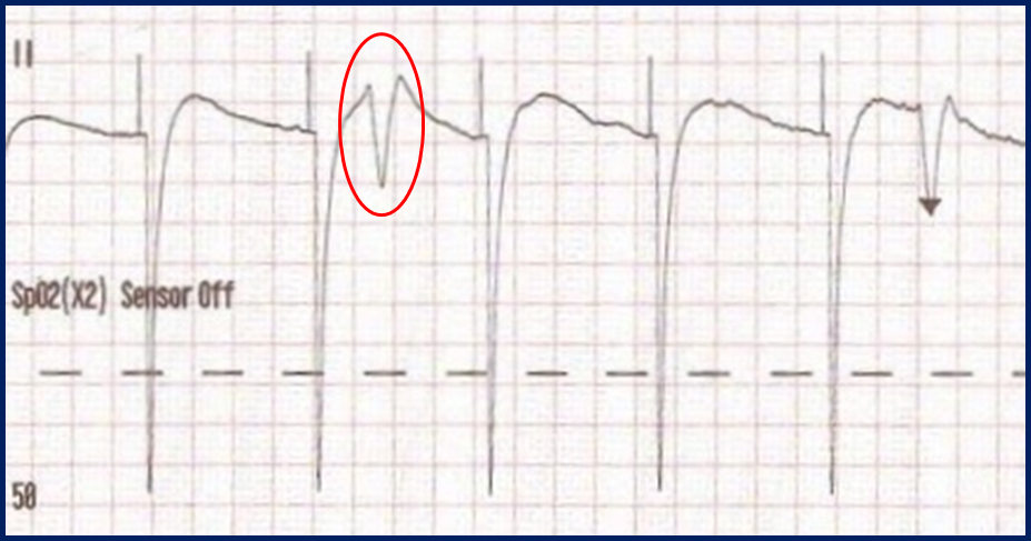
Classic “false capture” with near-vertical down-stroke of the (phantom) QRS complexes, slightly curved return to the isoelectric line, and unimpressive T-waves.
Identifying True Electrical and Mechanical Capture
True electrical capture is identified by organized, uniform wide QRS complexes with a notable T-wave and ST segment. Look at the following example of true electrical capture on an ECG monitor:
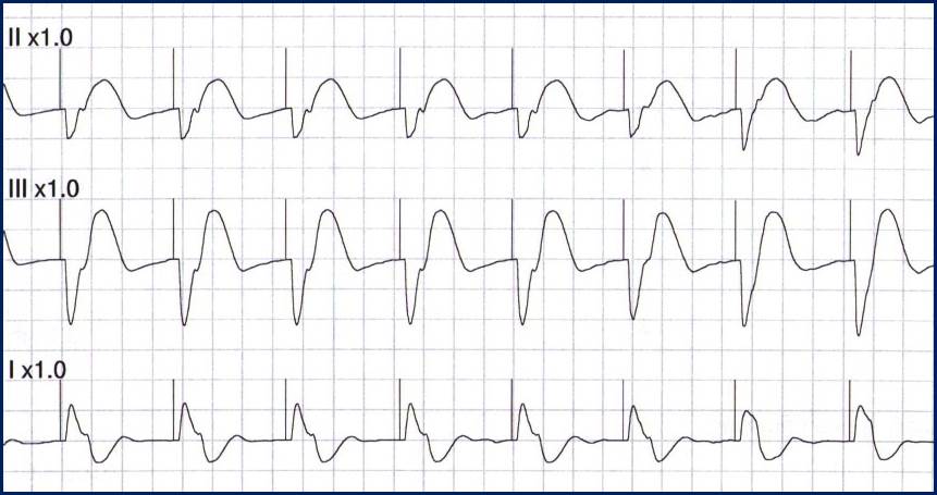
Transcutaneous pacing (TCP) with true electrical capture as evidenced by wide QRS complexes with tall, broad T-waves. Capture was achieved at 90 mA (ems12lead.com).
However, while this confirms electrical capture, you must also confirm mechanical capture to ensure adequate perfusion pressure for the patient. Otherwise, the possibility of false capture persists without confirming a therapeutic effect.
Tools to Confirm Mechanical Capture
In addition to electrical capture, mechanical capture should be confirmed via multiple methods:
- Palpable pulse (femoral preferred over carotid)
- Increase in blood pressure
- Improved pulse oximetry with a corresponding waveform
- Improved skin color and temperature (warm/pink)
- Improved level of consciousness
- Improvement in EtCO2
- Doppler ultrasound on a femoral or radial artery
Note that a palpable pulse alone is insufficient for determining adequate perfusion since ineffective transcutaneous pacing can still cause muscle jerks that mimic the feeling of a pulse at the site of palpation. This will create the same sensation for TCP with and without capture.
Common Reasons for Failure to Capture
One of the most common reasons for failure to capture with TCP is insufficient milliamperes (or current). If the current isn’t set high enough to overcome factors such as obesity or excessive chest hair, the current may never reach the heart for depolarization. Sometimes, the provider finds the initial threshold for true capture but then fails to leave a “buffer” increase of an extra 10 mA above the threshold to ensure that capture is not lost.
Another reason for failure to capture is poor pad placement. As of present-day practice, pads should be placed in the anterior-posterior position with one over the left frontal chest and the other on the back directly posterior to the heart. In the former method, anterior-lateral pad positioning, the vector of current could easily miss a significant portion of the ventricle for depolarization, thus requiring a higher level of current to obtain capture.
Practical Tips for Successful Transcutaneous Pacing
When performing TCP, remember these key tips to prevent false capture:
- Perform, but do not rely on, a pulse check (do not be fooled by skeletal muscle contraction) whenever determining capture.
- Use multiple modalities to confirm mechanical capture whenever possible (SpO2, Doppler, capnography, or echo).
- Always set a pacer current “buffer” of 10mA’s above the original threshold for achieving capture.
- Know that the patient may become more alert whether capture is achieved or not.
- The most common reasons for “failure to capture” are insufficient milliamperes and poor pad placement.
As with many complex cardiac rhythms, remember that frequent cardiovascular assessment and monitoring are essential.
Key Clinical Takeaways for EMS and Emergency Providers
In summary, TCP is a lifesaving treatment modality that can be technically challenging, with false capture requiring early recognition to promote positive outcomes. ECG appearance alone is insufficient to confirm true capture in the patient, and multiple methods should be used to ensure cardiovascular perfusion in the patient.
When life is measured in seconds, a solid foundation in managing complex cardiac rhythms, whether pacing or otherwise, is essential to promote positive patient outcomes. ACLS Training and BLS Training courses provide a fast track to clinical mastery that fosters confidence in managing life-threatening cardiac issues. Formalize your expertise by signing up for a certification or recertification course today!
References
Abella BS, Aziz K, Berg KM, et al. 2025 American Heart Association and American Red Cross Guidelines for Cardiopulmonary Resuscitation and Emergency Cardiovascular Care. Circulation. 2025;152(16_suppl_1). doi:10.1161/CIR.0000000000001376
Kimbrell, J., Kreinbrook, J., Poke, D., Kalosza, B., Geldner, J., Shekhar, A. C., … Vega, J. (2024). False Electrical Capture in Prehospital Transcutaneous Pacing by Paramedics: A Case Series. Prehospital Emergency Care, 28(7), 928–936. https://doi.org/10.1080/10903127.2024.2321287
Sun Y, Zhang J, Wang X, et al. Abstract Sun1205: Transcutaneous pacing in the prehospital setting – challenges, limitations and opportunities for improvement. Circulation. 2025;152(Suppl_3):ASun1205. doi:10.1161/circ.152.suppl_3.Sun1205
Swerdlow CD, Wang PJ. Temporary cardiac pacing. In: Post TW, ed. UpToDate. Waltham, MA: UpToDate Inc. Updated September 23, 2025. Accessed April 2, 2026. https://www.uptodate.com/contents/temporary-cardiac-pacing