ACLS Approach to Unstable Bradycardia: Atropine, Pacing, and Clinical Pitfalls
Treatment for unstable bradycardia can be daunting for clinicians as it is a time-sensitive, high-stakes condition with risks for hemodynamic compromise. Nonetheless, swift identification and response using ACLS principles can prevent complications, with initial interventions including IV medications like atropine or transcutaneous pacing (TCP), skills that are often reinforced through online ACLS training and certification . We will review one such case below.
Table of Contents
- What Is Unstable Bradycardia?
- Key Questions to Ask When Evaluating Bradycardia
- Signs of Hemodynamic Instability in Bradycardia
- Case Presentation: Severe Symptomatic Bradycardia
- ECG Findings in High-Degree AV Block
- Initial Management of Unstable Bradycardia
- Atropine in Bradycardia: When It Helps and When It Does Not
- Transcutaneous Pacing (TCP) as a Temporary Measure
- Rhythm Conversion and Clinical Improvement
- Reversible Causes of Bradycardia You Must Consider
- Definitive Management of High Degree AV Block
- Clinical Takeaways for Bradycardia Management
- Strengthening Bradycardia Management Skills With ACLS Training
What Is Unstable Bradycardia?
Bradycardia is defined as a resting heart rate below 60 beats per minute. It is considered asymptomatic when the patient is adequately perfused, but symptomatic bradycardia is marked by signs of hypoperfusion including dizziness, syncope or fatigue. Importantly, there is no single heart rate threshold that leads to the development of symptoms; rather, treatment is guided by the patient’s symptomatology and hemodynamic status than heart rate alone.
Key Questions to Ask When Evaluating Bradycardia
When considering bradycardia management, 3 initial questions should be answered:
- Is the patient bradycardic?
- Is the patient symptomatic?
- Is the patient symptomatic from the bradycardia?
If all of these have been answered with a YES, determine the patient’s hemodynamic status.
Signs of Hemodynamic Instability in Bradycardia
Signs of hemodynamic instability include:
- Hypotension
- Altered mental status
- Ischemic chest discomfort
- Acute pulmonary edema
- Signs of shock or hypoperfusion (pale, cool to touch, thready pulse, low urine output)
Case Presentation: Severe Symptomatic Bradycardia
Consider the following scenario:
A 75-year-old male experienced a syncopal episode. The event was witnessed by family members who contacted 9-1-1. On arrival of EMS personnel, the patient appears acutely ill. He is pale, diaphoretic and cool to touch. He states that he is feeling lightheaded and weak.
Medical History
- Hypertension
- Hyperlipidemia
- Gout
- Bilateral knee replacement
- Left bundle branch block
The family reports the patient is seeing a cardiologist and is scheduled for pacemaker implantation in 3 weeks due to previous episodes of symptomatic bradycardia.
Medications
- Zocor (Simvastatin)
- Lopressor (Metoprolol)
- Aloprim (Allopurinol)
- Multi-vitamins
Vital Signs
- RR: 20
- HR: 20
- BP: 80/48 mm Hg
- SpO2: 93% on room air
- Capillary blood glucose: 118 mg/dL
Breath sounds are clear bilaterally.
The patient is placed on O2 via nasal cannula at 2 LPM with ETCO2 of 16 mmHg.
ECG Findings in High-Degree AV Block
Cardiac monitoring is established and the following 12 lead ECG is obtained.
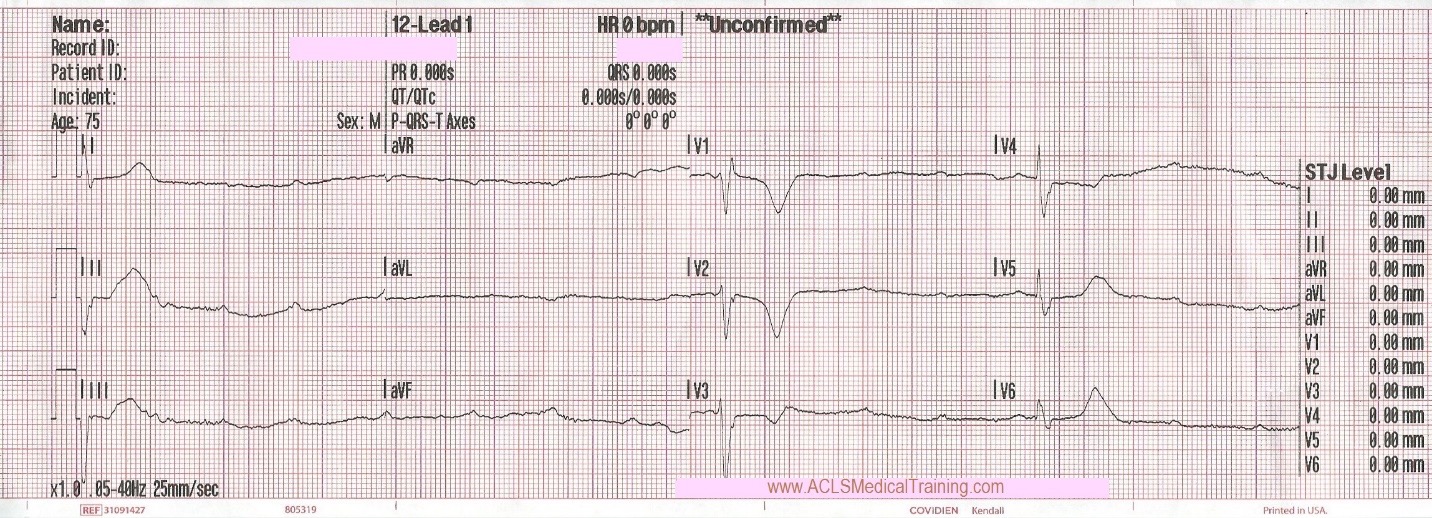
High degree AV block with heart rate less than 20.
A high degree AV block is form of second degree block where at least 3 P waves occur before a QRS (P:QRS ratio of 3:1 or higher). This is in contrast to a 3rd degree block where P and QRS waves “fire” without respect to each other (known as AV Dissociation).
An escape rhythm, as is pictured in the unstable bradycardia case study above, is a compensatory mechanism that occurs when the higher cardiac node signals are “blocked” from reaching the lower portions of the heart. When the SA (atrial) node is blocked, the AV “junction” takes over with a junctional escape rhythm (typically 40-60bpm with a narrow QRS); if the AV node fails, then the ventricle takes over with a ventricular escape rhythm (typically 20-40 bpm with wider QRS complexes). When a block occurs below the AV node, known as an “infranodal block”, the slower conduction of the ventricles causes a wider QRS than the higher nodal conduction of the AV node and Bundle of His. Thus, wide complex escape rhythms signal a block below the AV node with reliance on the ventricles.
Initial Management of Unstable Bradycardia
In addition to heart rate, patients with unstable bradycardia require blood pressure monitoring to assess for hypotension and IV access for administering medications or fluid resuscitation. Additionally, oxygen saturation must be monitored to assess for hypoxia. Supplemental oxygen may be given even if the oxygen saturation is above 92% but below 100% to address any underlying hypoxemia which may be contributing to the arrhythmia. It is also critical that pacer pads be placed prophylactically in case of hemodynamic compromise.
Atropine in Bradycardia: When It Helps and When It Does Not
Atropine counteracts increased vagal tone (a known source of bradycardia) by binding to receptors in the SA and AV node, where vagal innervation is abundant, to improve conduction. However, since there is little to no vagal innervation below the AV node, Atropine is not believed to have a direct effect on bradycardic rhythms of ventricular origin. Thus, the decision for when to use atropine in bradycardia will depend on the presence of an infranodal AV block. These distinctions, including appropriate atropine dosing and when to escalate to pacing or vasoactive infusions, are emphasized in ACLS recertification training to ensure clinicians stay aligned with current guidelines.
For clinicians asking, “does atropine work in third degree AV block?”, the 2018 ACC/AHA/HRS Guideline on the Evaluation and Management of Patients with Bradycardia caution:
“Atrioventricular block within or below the His bundle… will not respond to Atropine but will sometimes improve with catecholamines… Atropine is unlikely to improve atrioventricular block at the His bundle or His-Purkinje level and isolated reports have suggested occasional worsened atrioventricular conduction and/or hemodynamic compromise in such patients. For this reason, Atropine should be used judiciously in patients with atrioventricular block and wide QRS complexes that suggest the presence of significant His Purkinje disease.”
Transcutaneous Pacing (TCP) as a Temporary Measure
Regarding transcutaneous pacing indications, ACLS guidelines for bradycardia are informed by the 2025 AHA CPR and ECC Guidelines which advise:
“When symptomatic bradycardia is refractory to medical management, cardiac pacing is a reasonable alternative. Transcutaneous pacing is noninvasive, can be performed quickly at bedside, and is typically used as a bridge to placement of a temporary transvenous pacing catheter.”
With TCP, the clinician must ensure electrical capture by confirming a wide QRS after each pacer spike on the ECG monitor with hemodynamic improvement. Once capture is achieved, the clinician should increase the current by an additional 5-10 mA to ensure reliable capture.
Transcutaneous pacing (TCP) is notoriously painful and often requires the patient to be sedated until they can be bridged to transvenous pacing. Additionally, TCP is inefficient, requiring high voltage “shocks” through the chest wall to pace the heart. For these reasons, it is meant to be a temporary bridge therapy until the patient can undergo placement of a temporary transvenous pacemaker or a permanent implanted pacemaker.
In addition to TCP, the updated 2025 AHA CPR and ECC Guidelines also give increased weight to adrenergic alternatives stating:
“Dopamine infusion can also increase heart rate. There are limited studies comparing medications to transcutaneous pacing for the treatment of bradycardia. A randomized feasibility study in patients failing atropine compared dopamine to transcutaneous pacing and found no difference in survival to discharge. Whether to trial transcutaneous pacing, epinephrine, dopamine, or other vasoactive agent will likely, therefore, depend on clinician experience and resources available.”
Rhythm Conversion and Clinical Improvement
Let’s return to our scenario:
Now that the patient has been identified as having unstable bradycardia, defibrillation pads are placed prophylactically, IV access is obtained and 250 ml of normal saline is administered en route to the Emergency Department 4 minutes away.
Upon arrival blood samples are obtained and the following 12 lead ECG is obtained.
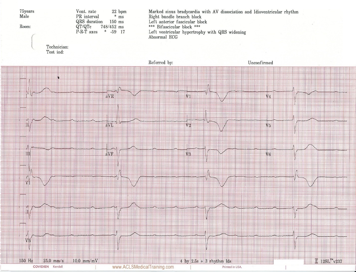
3rd Degree AV Block. The escape rhythm shows a wide QRS with bifascicular morphology (RBBB morphology with left axis deviation). It is likely ventricular in origin.
The patient’s level of consciousness deteriorates and he responds only to painful stimuli.
1 mg atropine is administered rapid IV push followed by 10 ml saline flush.
After 1 minute, transcutaneous pacing is initiated with no electrical capture up to 90 mA. Transcutaneous pacing is discontinued by the arriving cardiologist who requests vasopressors.
Prior to vasopressors being administered a change is noted on the cardiac monitor and another 12 lead ECG is obtained.
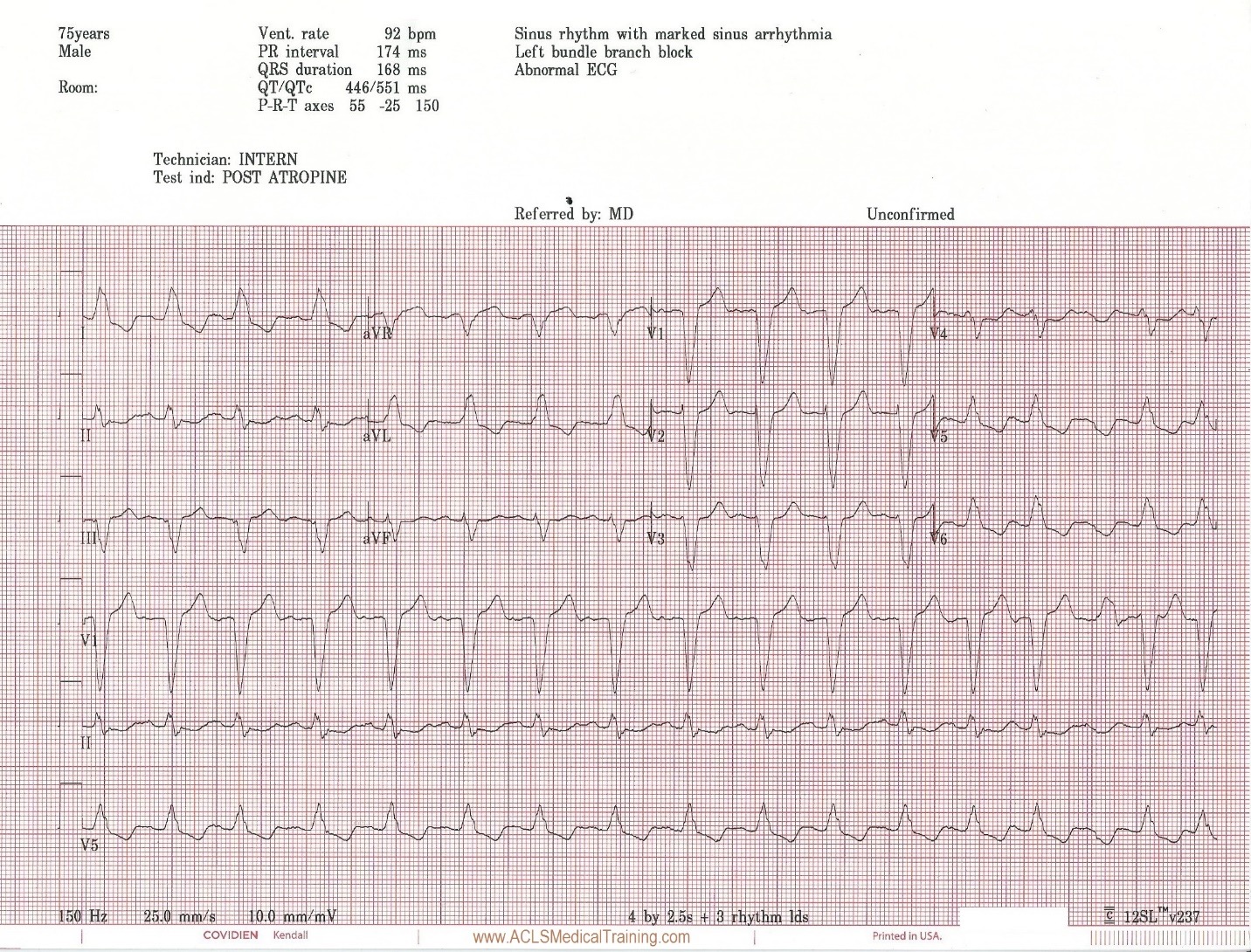
The bradycardia has resolved and the heart rate is now sinus rhythm at 92 bpm. There is a left bundle branch block which is consistent with the patient’s known medical history.
The patient now reports feeling better with improved skin color and blood pressure. The patient is taken to cardiac cath lab for angiography and a permanent pacemaker. The procedure was successful and he was placed in the cardiac step-down unit for further observation.
In this unstable bradycardia case study, transcutaneous pacing was not successful, but the clinicians who were caring for this patient realized that they had not achieved capture, which is not always the case !
Fortunately, the patient spontaneously converted into a perfusing rhythm. Possible reasons for conversion back into sinus rhythm could be the atropine which was administered, sympathetic stimulation from attempted transcutaneous pacing, or it could be a coincidence. These types of scenarios are susceptible to the “post hoc ergo propter hoc” fallacy (after this, therefore because of this).
Reversible Causes of Bradycardia You Must Consider
A common error when treating patients with bradycardia is a rush to drug or electrical therapy prior to identifying reversible causes. Remember, Hs and Ts aren’t just for asystole and PEA!
Reversible causes of unstable bradycardia may include:
- Hypoxia
- Medication effects (e.g. Calcium-Channel blockers, Beta-Blockers, Digoxin)
- Myocardial ischemia
- Electrolyte disturbance (e.g. Hyperkalemia)
- Acidosis
- Hypothermia
Of these, hypoxemia should be rapidly identified and treated, but other conditions like hyperkalemia can also cause bradycardia. In the case of hyperkalemia, there is little to lose and much to gain from giving a patient calcium gluconate or calcium chloride prior to pacing. To quote Stephen Smith, M.D.: “The treatment [of hyperkalemia with calcium] is benign and cheap. How many life-threatening diseases can you treat benignly and cheaply?”
Definitive Management of High Degree AV Block
If unstable bradycardia persists despite medications and addressing potential reversible causes, TCP should be initiated but replaced with transvenous pacing as soon as possible to bridge the patient until definitive treatment can be performed (e.g. permanent pacemaker placement).
For the patient with unstable bradycardia, cardiology consultation is recommended in order to guide treatment as there is significant latitude for clinical judgment in choosing between types of medical management (Atropine, Dopamine, Epinephrine) or pacing (whether transcutaneous or transvenous) based on patient presentation and hospital resources.
Clinical Takeaways for Bradycardia Management
As in many conditions, “treat the patient, not the monitor” for patients with bradycardia. As soon as the patient develops signs of cardiopulmonary compromise, the instability must be addressed per ACLS protocol. In addition, TCP must not be delayed when indicated since complications from hypoperfusion can escalate quickly, and the provider should consider reversible causes as soon as bradycardia presents.
Strengthening Bradycardia Management Skills With ACLS Training
ACLS bradycardia algorithms require familiarity and rapid pattern recognition due to the risk for hemodynamic compromise. In the case above, adherence to ACLS principles prevented prolonged hypoperfusion and further clinical deterioration.
Confidence and accuracy in recognizing complex cardiac rhythms begin with strong foundational training and ongoing education. Whether you are obtaining initial certification or renewing your credentials, structured review of emergency cardiovascular care protocols improves both clinician confidence and patient outcomes.
We offer comprehensive online training options to support every stage of your professional development:
- ACLS Certification : Initial Advanced Cardiovascular Life Support training covering cardiac arrest, bradycardia, tachycardia, stroke, and post–cardiac arrest care.
- ACLS Recertification : Streamlined renewal training focused on updated guidelines, algorithm review, and case-based scenarios.
- BLS Certification : Foundational Basic Life Support training in CPR, AED use, and early recognition of cardiopulmonary emergencies.
- PALS Certification : Pediatric Advanced Life Support training tailored to infant and child cardiac and respiratory emergencies.
Advance your knowledge, sharpen your clinical decision-making skills, and stay prepared for high-stakes cardiac events through online certification and recertification training.
References
EMCrit Podcast 42: A phD in EKG with Steve Smith. EMCrit Podcast. 2011. Available at: https://emcrit.org/emcrit/phd-in-ekg/. Accessed November 26, 2017.
Kusumoto FM, Schoenfeld MH, Barrett C, et al. 2018 ACC/AHA/HRS Guideline on the Evaluation and Management of Patients With Bradycardia and Cardiac Conduction Delay: A Report of the American College of Cardiology/American Heart Association Task Force on Clinical Practice Guidelines and the Heart Rhythm Society. Journal of the American College of Cardiology. 2019;74(7):e51-e156. Available at: https://www.jacc.org/doi/10.1016/j.jacc.2018.10.044 . Accessed February 9, 2026.
Panchal AR, Berg KM, Kudenchuk PJ, et al. Part 9: Adult Advanced Life Support: 2025 American Heart Association Guidelines for Cardiopulmonary Resuscitation and Emergency Cardiovascular Care. Circulation. 2025;152(suppl 2). Available at: https://www.ahajournals.org/toc/circ/152/18_suppl_2 . Accessed February 9, 2026.