Treating Supraventricular Tachycardia with Adenosine
Adenosine is one of the primary medications used to treat certain types of supraventricular tachycardia (SVT). But when is it appropriate to use adenosine for SVT? This article will follow a real-world scenario to display the different facets of adenosine including how it works, appropriate usage, how much adenosine is appropriate, accurate ECG interpretation and important safety considerations, all of which are critical information for ACLS certification renewal.
Table of Contents
- What Is Supraventricular Tachycardia?
- Initial Patient Assessment for SVT
- Identifying SVT on an ECG
- Initial Treatment for Stable SVT
- When Is Adenosine Used for SVT?
- Adenosine Dose for SVT
- ECG Changes During Adenosine Administration
- Confirming Successful Conversion to Sinus Rhythm
- When Adenosine May Not Be Appropriate
- Common Side Effects and Patient Monitoring
- What Happens If Adenosine Doesn't Work?
- Learn How to Treat Supraventricular Tachycardia with Adenosine
What Is Supraventricular Tachycardia?
Supraventricular Tachycardia (SVT) describes a group of abnormally accelerated cardiac rhythms that originate above the ventricles (hence, “supra-ventricular”), specifically above the Bundle of His. These rhythms are often characterized by narrow QRS complexes, though there are exceptions to this. SVT is also used by clinicians as a shorthand for paroxysmal supraventricular tachycardia (or PSVT) which is a type of tachycardia with abrupt onset and ending.
SVT typically develops via a reentry circuit where accessory pathways with differing conduction speeds and refractory periods perpetuate a self-sustaining loop of rapid electrical impulses. Thus, SVT encompasses a large category of arrhythmias with differing treatments, and adenosine is only appropriate for particular regular narrow-complex tachycardias. With any SVT in emergency and critical care settings, rapid rhythm identification is crucial due to the potential for conversion to deadly rhythms and a dangerous increase in cardiac demand.
Initial Patient Assessment for SVT
Consider the following scenario: EMS are dispatched to a home for a 59-year-old male complaining of palpitations, anxiousness, and shortness of breath. Symptoms began an hour prior with a remote history of a PSVT episode 10 years ago. The patient takes no medications and has no known drug allergies.
Upon arrival, the patient looks anxious but is oriented x4. Vital signs are as follows:
- HR: 213
- NIBP: 128/92
- RR: 22
- Temp: 98.5 F
- SpO2: 96% on RA
- BGL: 91
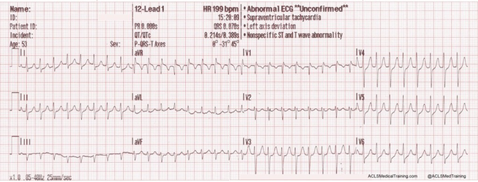
In this scenario, it is crucial to assess patient stability as the first priority before determining treatment as signs of poor perfusion require rapid correction. EMS personnel obtain a 12-lead ECG which shows the following:
A brief rhythm strip is also recorded:
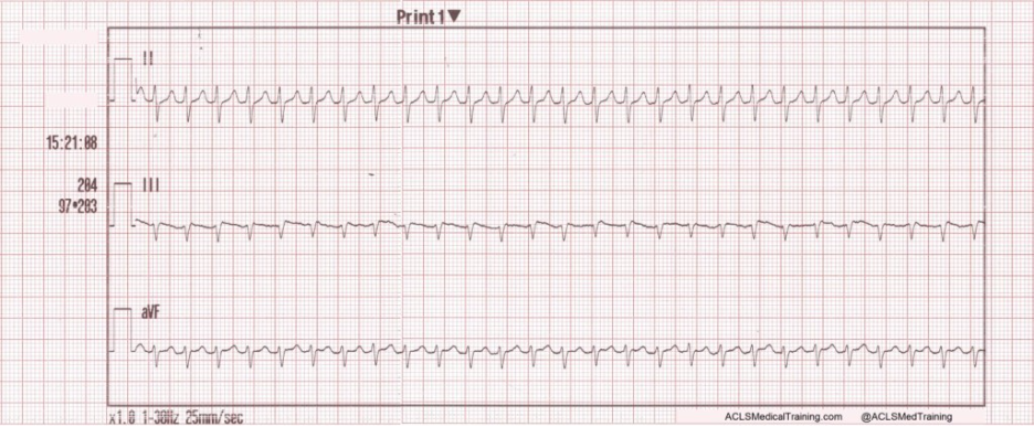
The heart rate found on the ECG is approximately 200 beats per minute, appearing as a regular narrow complex tachycardia. Such a type of patient can be encountered by clinicians, whether
Identifying SVT on an ECG
SVT is commonly seen as a regular, narrow QRS complex at a rapid rate ranging from 150 to 250 bpm. Clinicians cannot absolutely rule SVT in or out based on ECG alone, but some findings that indicate SVT include an abrupt start and stop, a constant rate that does not change with activity or stress, and often narrow QRS complexes (though this is not always the case).
SVT typically has a regular rhythm with a narrow QRS width of <120 milliseconds. Due to the rapid heart rate, P waves are often “hidden” within the ECG tracing and cannot be seen unless the rate is slowed via vagal maneuvers or intravenous adenosine. Clinical context is important for ruling SVT in or out, including sudden onset and offset, a feeling of palpitations, and triggers including caffeine, stress, and response to vagal maneuvers.
Initial Treatment for Stable SVT
For patients with stable SVT, management starts with vagal maneuvers when the patient can follow instructions and be monitored. Vagal maneuvers are favored because they slow AV nodal conduction, have the ability to terminate reentrant SVT, and can also help clarify the rhythm by making atrial activity (P waves) more visible. During vagal maneuvers, continuous cardiac rhythm monitoring is essential, ideally with a 12-lead ECG when possible.
The modified Valsalva maneuver is a viable and effective treatment for SVT with a successful conversion rate of over 40% compared to the standard Valsalva, per the REVERT trial. With the modified version, clinicians should have the patient sit in a semi-recumbent position and produce artificial strain by blowing into an object such as a small plastic syringe for 15 seconds, followed by supine positioning for 15 seconds with passive leg raise at a 45-degree angle (see this video example of the procedure). Following any maneuvers, patient assessment is critical to confirm hemodynamic stability and potential conversion out of SVT prior to any attempts at pharmacologic intervention.
When Is Adenosine Used for SVT?
Adenosine is typically considered for stable patients with a regular narrow-complex tachycardia when vagal maneuvers have failed to terminate the rhythm. Before moving to medication, clinicians may attempt techniques such as the modified Valsalva maneuver; see a video example of the procedure for a demonstration.
Before administering adenosine, clinicians should carefully assess the patient's hemodynamic stability and evaluate the rhythm, including whether the QRS complex is narrow or wide and whether the rhythm is regular or irregular. Adenosine may also be considered in select stable patients with a regular, monomorphic wide-complex tachycardia when the underlying rhythm is uncertain.
Adenosine should not be used indiscriminately for all tachyarrhythmias. It should be avoided in irregular or polymorphic wide-complex tachycardias, and clinicians should consider important contraindications and precautions before administration. If the patient is hemodynamically unstable, synchronized cardioversion is generally the priority, although adenosine may be considered in certain regular narrow-complex rhythms while preparations for cardioversion are underway.
Adenosine Dose for SVT
So, what is the proper dosage and administration method for adenosine in SVT? Adenosine is administered via rapid IV push followed by a saline flush. Rapid administration is important because efficacy depends on rapid delivery to the heart.
It’s important to note that the following the first dose of adenosine for SVT in adults and pediatrics, the second dose is doubled. The adenosine dosage guidelines for adults and children with a regular and monomorphic SVT are as follows:
| Adult | Pediatric | |
| 1st Dose | 6mg IV Push | 0.1mg/kg IV Push (Max: 6mg) |
| 2nd Dose | 12mg IV Push (if required) | 0.2mg/kg (Max: 12mg) |
Based on 2025 Guidelines from the American Heart Association and American Academy of Pediatrics
It is critical that clinicians have familiarity and ready access to dosing information for adenosine as well as other medications which are covered in our bundled BLS, ACLS and PALS courses.
ECG Changes During Adenosine Administration
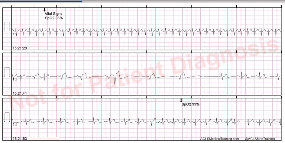
Upon administering adenosine, clinicians can expect to see one of several findings:
- Transient AV block: Because adenosine slows conduction through the AV node, a transient pause in ventricular activity (despite atrial pacing) is expected prior to successful conversion to a normal sinus rhythm.
- Rhythm pause or slowing: A rhythm pause appears as a brief flat line and can be a scary but expected finding so long as it doesn’t persist. Rhythm slowing will bring the heart rate down significantly, and perhaps even to a bradycardic rate, but this too is brief and should be followed by a return to normal sinus rhythm.
- Successful conversion: Whether a transient block, rhythm pause or significant slowing is seen, successful conversion looks like a return to normal sinus rhythm with a regular rate and visible P waves.
See below for examples of the above intervening ECG findings:
Confirming Successful Conversion to Sinus Rhythm
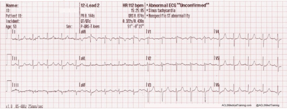
Confirmation of SVT resolution is accomplished by the reappearance of P waves with a clear sinus rhythm tracing and improvement of symptoms including palpitations, shortness of breath, dizziness, nausea and anxiety. However, even with these reassuring signs, a repeat 12-lead ECG should always be performed to confirm SVT resolution and to investigate any underlying rhythms or conditions that are contributing to the presence of SVT.
The following ECG strip demonstrates complete SVT resolution and conversion back to sinus rhythm after adenosine administration:
When Adenosine May Not Be Appropriate
Adenosine is contraindicated for irregular wide-complex tachycardias, atrial fibrillation with rapid ventricular response, atrial fibrillation with Wolff-Parkinson-White Syndrome, and polymorphic ventricular tachycardia as many of these irregular rhythms can precipitate dangerous conversion into ventricular fibrillation with hemodynamic collapse and possible death. Additionally, adenosine is contraindicated in hemodynamically unstable patients as well as patients with asthma due to the risk for severe bronchospasm. In almost all of these scenarios, synchronized cardioversion is the first-line treatment as it does not carry the same risks of conversion into ventricular fibrillation.
Common Side Effects and Patient Monitoring
The most common side effects that patients experience after receiving adenosine are caused by the transient AV block which the medication produces. These side effects include:
- Facial flushing
- Shortness of breath
- Palpitations
- Chest pain
- Lightheadedness
Patients should be monitored with 12-lead ECG monitoring before, during and after adenosine administration. The patient should be placed supine with blood pressure measurements taken at least every 5 minutes initially. After administration, monitoring should continue until the patient’s rhythm and hemodynamics have clearly returned to baseline.
What Happens If Adenosine Doesn’t Work?
Why does adenosine sometimes fail? Most often, the drug is not efficacious because the problematic rhythm is not an AV-node dependent reentry SVT, the drug did not reach the central circulation fast enough, or the patient has personal factors that reduce adenosine’s effect.
If adenosine fails, the clinician should ensure that the patient’s rhythm is one amenable to the drug and check their administration technique to see if they delivered the drug quickly enough with a saline flush. A second adenosine dose that is doubled to 12mg may be given if no other explanation can be given for adenosine’s failure, and thereafter other medications may be tried (Beta blocker / Calcium channel blocker) as well as synchronized cardioversion for hemodynamically unstable patients.
Learn How to Treat Supraventricular Tachycardia with Adenosine
In summary, it is critical that clinicians recognize SVT quickly via ECG monitoring to determine QRS width and regularity. Use adenosine only when a regular monomorphic SVT is apparent and avoid giving it with rhythms such as irregular wide-complex tachycardias. Adenosine should only be used for stable patients placed in a supine position with constant ECG monitoring as well as frequent blood pressure readings before, during, and after administration to ensure stability and conversion back to a normal sinus rhythm.
When it comes to addressing dangerous cardiac rhythms, clinicians need a foundation that allows them to act safely and decisively. ACLS Medical Training offers top-of-class online medical training courses for healthcare professionals looking to master cardiac rhythm concepts and helps you save money via combination courses such as the ACLS and PALS training bundle. ACLS Medical Training also offers fundamental courses including a BLS recertification course. Take charge of your education to better serve your patients by registering for a course on our site today!
References
Appelboam A, Reuben A, Mann C, et al; REVERT trial collaborators. Postural modification to the standard Valsalva manoeuvre for emergency treatment of supraventricular tachycardias (REVERT): a randomised controlled trial. Lancet. 2015;386(10005):1747-1753. doi:10.1016/S0140-6736(15)61485-4
Nasir M, Sturts A, Sturts A. Common types of supraventricular tachycardia: diagnosis and management. Am Fam Physician. 2023;107(6):631-641.
Wigginton JG, Agarwal S, Bartos JA, et al. Part 9: Adult Advanced Life Support: 2025 American Heart Association Guidelines for Cardiopulmonary Resuscitation and Emergency Cardiovascular Care. Circulation. 2025;152(16_suppl_2):S538-S577. doi:10.1161/CIR.0000000000001376