Author Archives: Jameson McIntosh

Mixed Overdose and Na-Channel Blockade

It’s 06:30 when EMS is called to an inner-city apartment for an 18-year-old male having a seizure. After gaining entry into the building, Paramedics and First Responders trudge up two flights of stairs and down a narrow, dimly lit hallway, until they find an open door into a dark two-bedroom apartment.  There are three people in the living room, all about 18-years-old, and one of them is lying awkwardly on the floor, propped up against the side of the couch. There is evidence of extensive alcohol and drug consumption littering the scene.

It proves difficult to obtain a complete patient history, since bystanders on scene are still somewhat inebriated, and they remain apprehensive about cooperating with emergency responders. However, the story gathered is as follows.

The three of them were up all night partying, drinking, and ingesting multiple illicit substances. It is reported that the patient was witnessed to have ingested copious amounts of cocaine, “DXM” (Dextromethorphan, or ‘cough syrup’), marijuana, nicotine, and alcohol. Precise amounts are vague, and it’s possible that there may have been even more ingestions that were not reported. The party ended, and they all went to sleep, until one of the party-goers found the patient seizing on the floor at about 06:30 this morning. It’s unknown how long the patient was down prior to being found.

The patient’s medical history is unknown, though it is believed that he is generally a “pretty healthy guy”.

The patient is unresponsive to any stimuli, has a weak and agonal respiratory effort, and a faint and slow carotid pulse. His general appearance is poor, with significant pallor and central cyanosis noted, though he is hot to the touch.

Crews began administering high-quality 2-person BVM ventilation, utilizing a jaw thrust while inserting an OPA, and positioning the patient with padding behind the head to support a proper ear-to-sternal-notch alignment. Intravenous access, fluid resuscitation, reoxygenation, and basic cardiac monitoring is being maintained while extrication from the apartment is coordinated.

His initial vital signs are as follows:

HR – 60/min, and increasing to 100/min once oxygenation and ventilation is administered
RR – 6/minute and ineffective
SpO2 Initially <50%, improving to 83% with oxygenation and ventilation
NIBP – 66/34 [44]
Pupils – 6mm bilaterally, extremely sluggish
BGL – 5.0 mmol/L (90 mg/dl)
Temp – 38.5 Celsius (101.3 F)

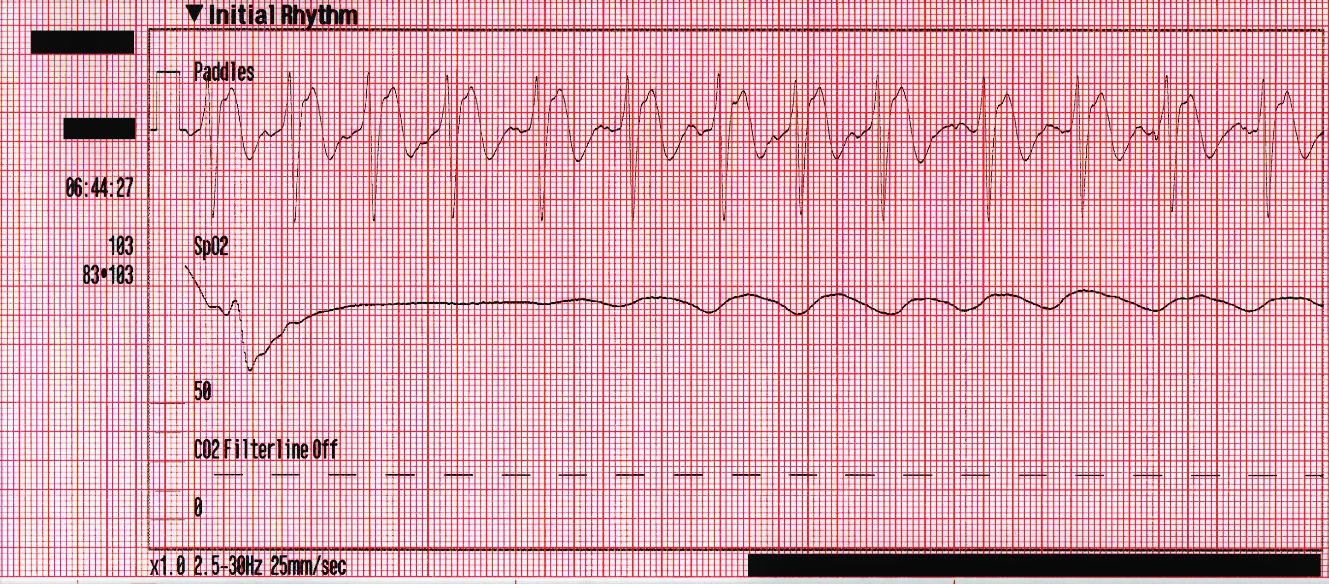
The following rhythm is observed on the monitor. Shortly later, the patient has a generalized seizure which lasts approximately 5 minutes.

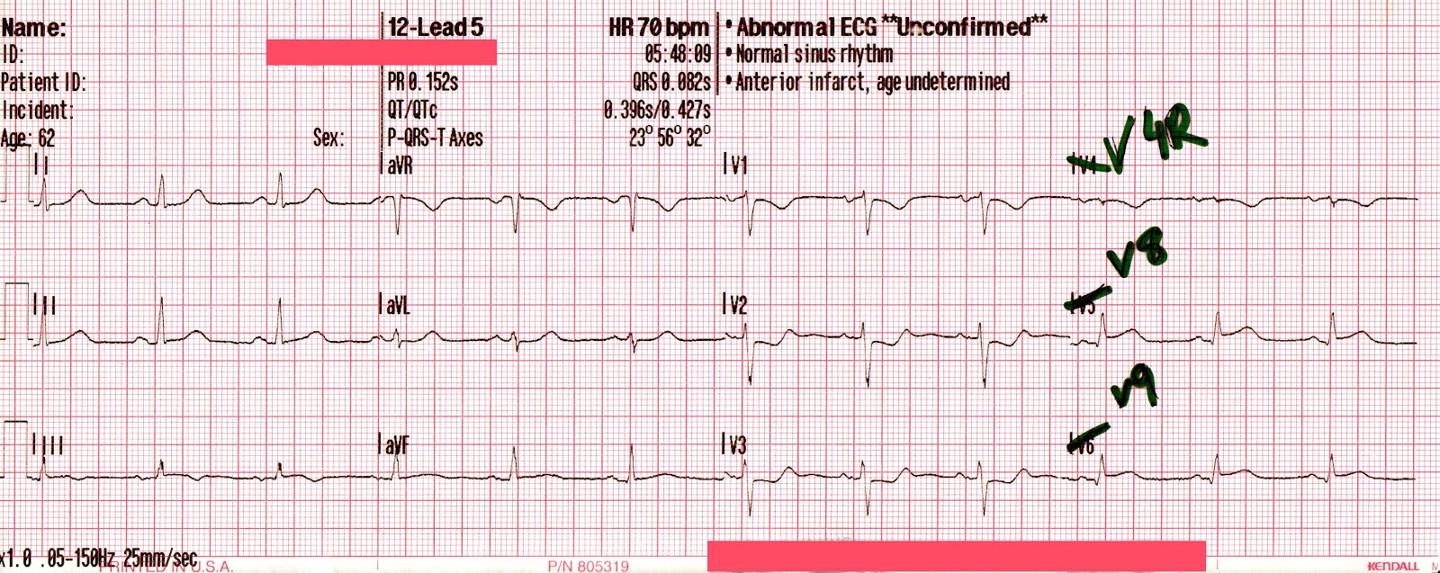
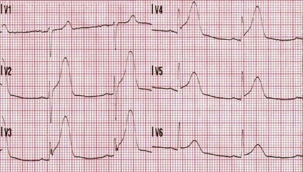
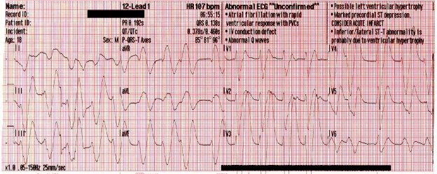
Due to access limitations, the stretcher could not be brought inside to the patient, and so the team of paramedics, police officers and firefighters worked together to move the patient to the ambulance via a device not unlike a large tarp with handles. Once in the ambulance, passive cooling is initiated, and a supraglottic airway is placed. His blood pressure has improved to 87/44 [59], his SpO2 remains in the mid-80’s, and his initial ETCO2 is 86mmHg. The following 12-lead is acquired:

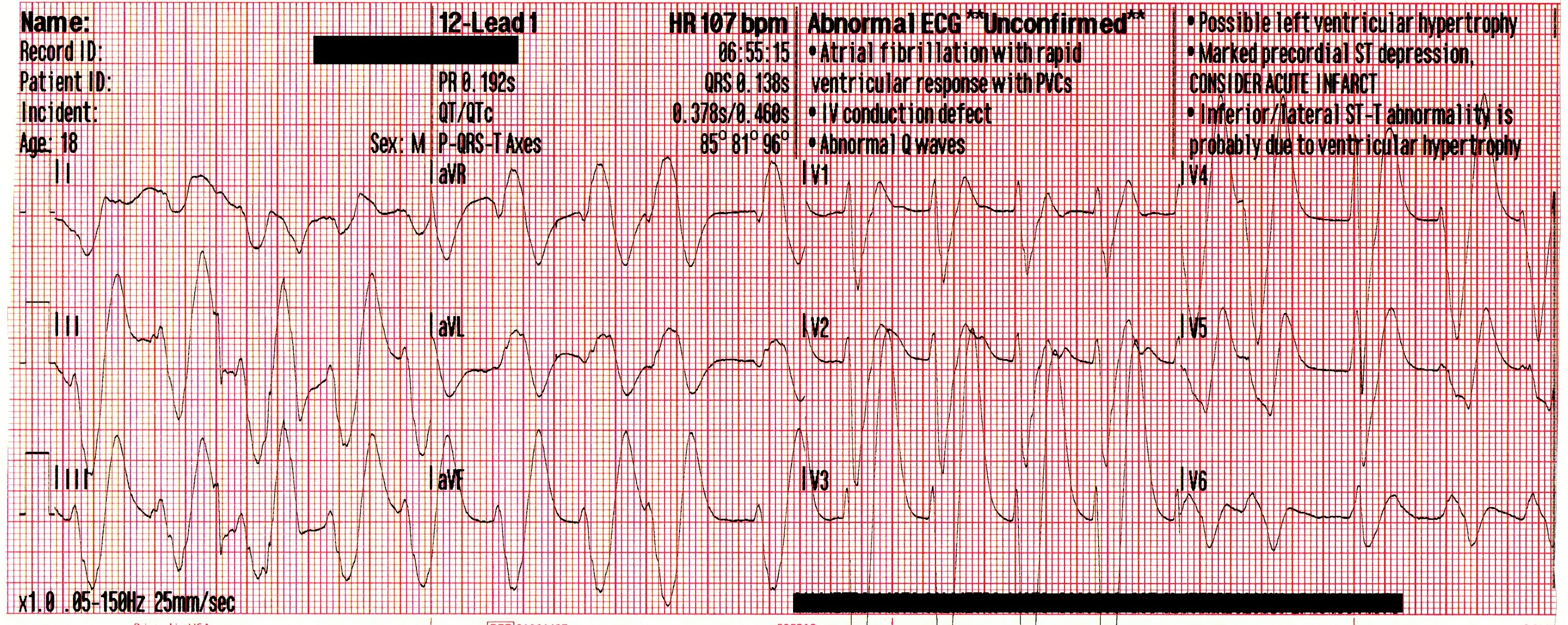
A significant wide-complex tachycardia that is irregularly irregular, with an extreme right axis deviation and a massive terminal R-wave in aVR measuring 10mm. Given the patient’s suspected ingestions and current clinical condition, this ECG should be considered pathognomonic for severe sodium-channel blockade being complicated by extreme acidosis.

Paramedics identified this arrhythmia to most-likely be a complication of the cocaine toxicity, and treatment was aimed at hyperventilation and administration of intravenous sodium bicarbonate (NaHCO3) to correct the acidosis.

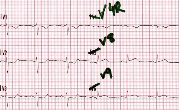
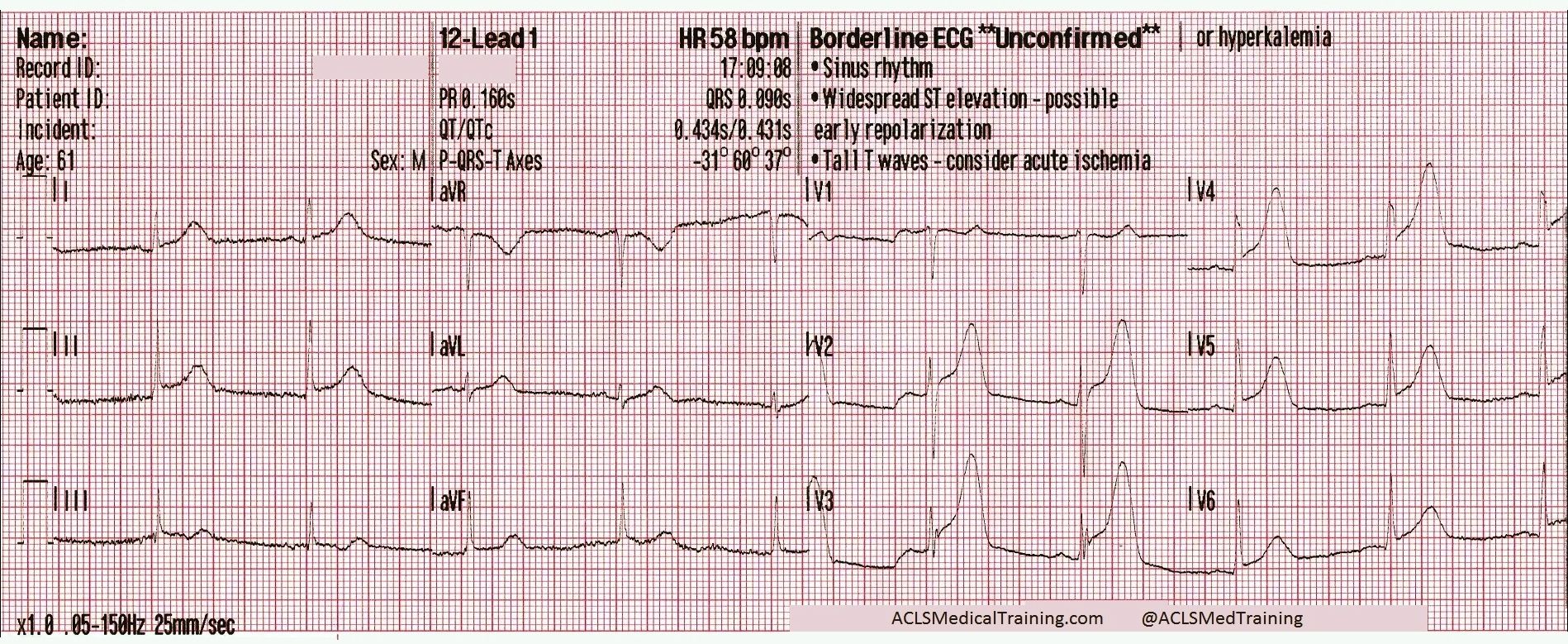
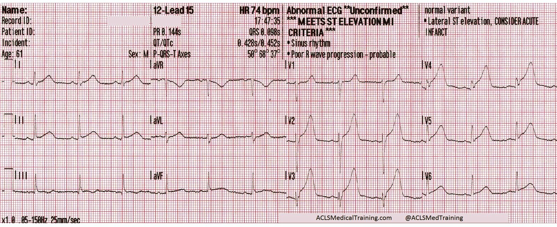
The following 12-lead was recorded following the administration of one amp of 50mEq of NaHCO3 with ongoing attempts at hyperventilation.

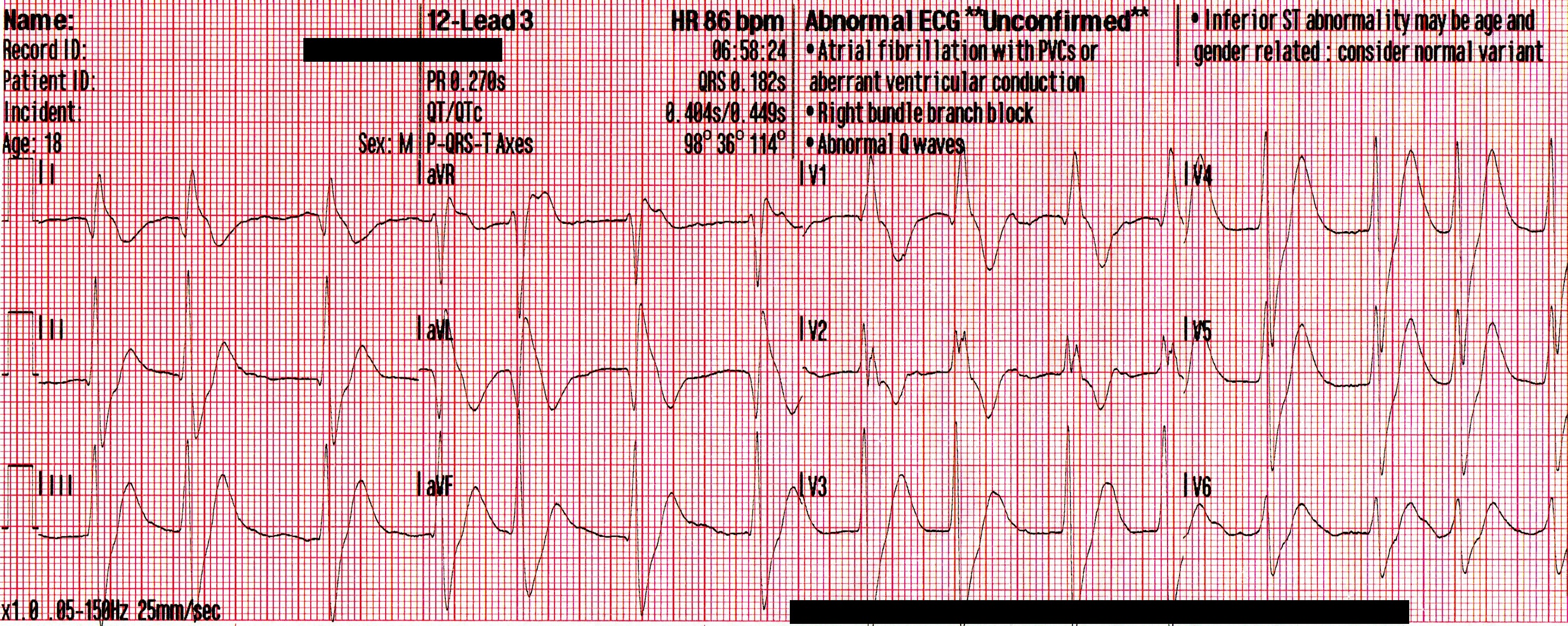
An irregularly-irregular wide-complex rhythm, with an apparent RBBB pattern and peaked T-waves reminiscent of hyperkalemia. This is an improvement, but there are still signs of significant sodium channel blockade.

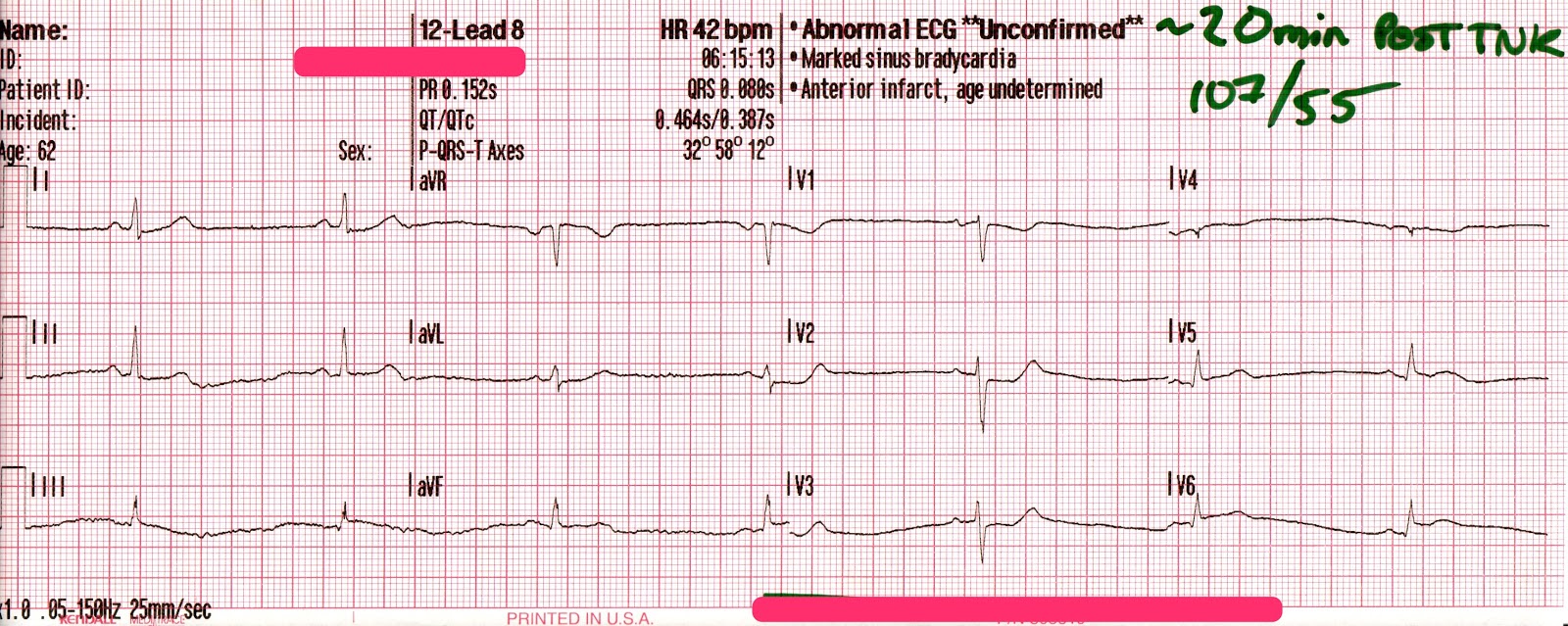
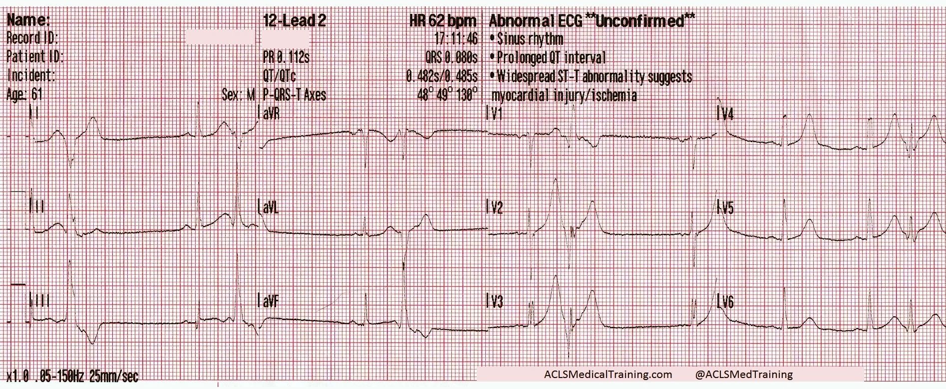
The patient’s SpO2 improved to 100%, his blood pressure remained 85/35 [55], and despite being ventilated at a rate of 30/minute his ETCO2 remained 86mmHg. Another 50mEq of NaHCO3 is administered, and the following 12-lead is acquired:

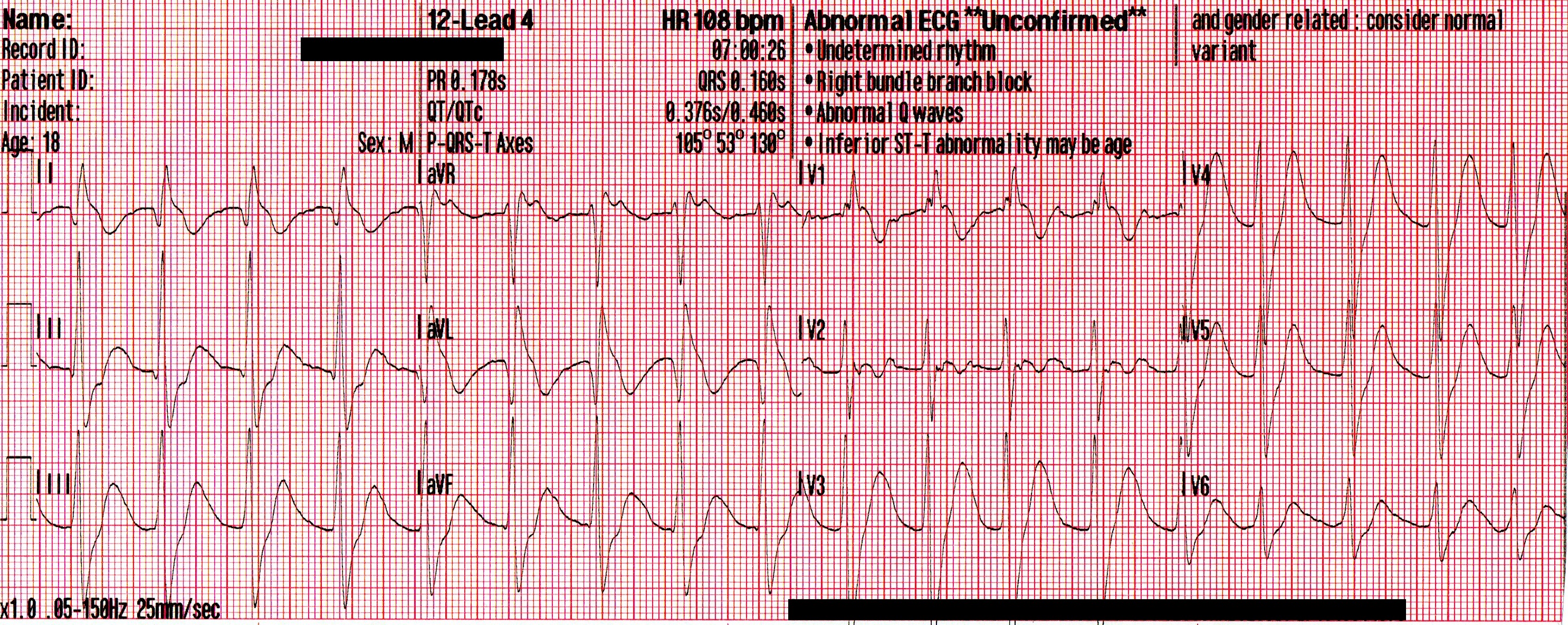
A regular, wide-complex rhythm, with a similar QRS morphology to the previous 12-lead. The QRS is gradually narrowing, but remains pathological.

Conclusion

The crew arrived at the hospital shortly after the second amp of NaHCO3 was given. The ED staff continued administering subsequent doses of NaHCO3 , a peripheral vasopressor (norepinephrine) was initiated, and he was intubated and placed on a ventilator. Initial arterial blood gases revealed a pH of <6.8, pCO2 of >100mmHg, and a lactate of >20mmol/L. He was sent for a CT-head, which revealed no obvious findings of hemorrhage or anoxic brain injury.

He was admitted to ICU, and his repeat ABG thirty minutes later revealed an improved pH of 7.28 and pCO2 of 48mmHg. Unfortunately, no further follow-up was made available to the author.

Discussion

The critically ill toxicology patient can present many unique challenges to prehospital and ED professionals alike. Obstacles often present themselves simultaneously, including airway compromise, cardiac dysrhythmias, and hemodynamic collapse. In an unconscious patient, this is further complicated by unknown co-ingestions, quantities, and comorbidities.

This patient’s presentation can likely be explained by the complex interaction between each of the substances that were ingested. Cocaine mixed with alcohol forms cocaethylene when metabolized by the liver; a substance that’s significantly more cardiotoxic, and possesses a half-life 3-5 times that of cocaine alone. Amongst it’s multiple mechanisms, it acts as a Class Ic sodium-channel blocker, which is represented on the ECG as a progressive widening of the QRS complexes, and the development of an extreme rightward axis in the frontal plane. These channel-toxic effects are amplified by increases in heart rate and decreases in pH – two elements that are found in spades for this young man.

The deleterious effects of the cocaethylene, combined with the ingestion of significant amounts of dextromethorphan; an antitussive and a NMDA-receptor antagonist;  would likely result in euphoria, tachycardia, hypertension, dissociation, a decreasing level of consciousness, and potentially severe serotonin syndrome. Hyperthermia, tachycardia, and a disrupted respiratory drive would lead to hypercapnia, worsening acidosis, and a decreased seizure threshold. Left unchecked, this would predictably spiral into a self-perpetuating loop, inevitably resulting in profound shock and hemodynamic collapse.

Treating a patient like this with intravenous sodium bicarbonate (NaHCO3) provides a multi-pronged attack. Following administration, there’s an rapid dissociation of NaHCO3 into Na + HCO3. The extra sodium acts to “overload” the sodium-channels blocked, while the bicarbonate acts as a buffer and binds with free hydrogen (H+) ions to form Carbonic Acid (H2CO3), which then dissociates into water and carbon dioxide, expressed as HCO3 + H H2CO3 H2O + CO2. This allows for respiratory correction of the acidosis, and the subsequent alkalinization of the blood helps to reduce the channel-toxic effects of the cocaine.  It should be noted, however, that this requires an increased rate of ventilation to ensure adequate elimination of the rising CO2 levels that will follow.

In a case as advanced as this one, where severe decompensated shock has developed, stabilization becomes a delicate and complex hurdle. Since our initial treatments are aimed at alkalinization of the blood to reduce cardiotoxicity, there is a resultant left-shift of the oxyhemoglobin dissociation curve, and that leads to a decreased ability for oxygen to offload from the hemoglobin at the level of the tissue beds. This could potentially hamper our attempts to correct the massive hypoxia that’s developed, and so management is usually targeted at a pH of no higher than 7.50-7.55.

Intubation of this patient would also prove delicate, since critical hypotension and acidosis would likely be worsened by the use of most induction agents or paralytics, forcing providers to classify this as a physiologically difficult airway. For this reason, airway management should likely be accomplished using a resuscitate-before-you-intubate approach. Fluid resuscitation should be well underway before RSI, push-dose pressors should be at the ready, and providers should be aware that there’s a high-likelihood of this patient requiring vasopressor support, despite receiving a 20ml/kg crystalloid bolus.

In conclusion, the critically ill mixed-overdose patient requires aggressive yet calculated emergency management from first responders and physicians alike. A clinical understanding of the pathophysiology, as well as the implications of each aspect of treatment, is vitally important in caring for each of these patients.

Further reading on the subject

Cocaine Overdose Presents with Wide Complex TachycardiaAlec Weir, M.D. ACLSMedicalTraining.com/Blog (2016)

Role of voltage-gated sodium, potassium and calcium channels in the development of cocaine-associated cardiac arrhythmiasMichael E. O’Leary & Jules C. Hancox. British Journal of Clinical Pharmacology (Oct 2009)

Current Concepts: The Serotonin SyndromeEdward W. Boyer M.D., Ph.D., Michael Shannon M.D., M.P.H. NEJM (2005)

Treatment of patients with cocaine-induced arrhythmias: bringing the bench to the bedsideRobert S Hoffman Br J Clin Pharmacol. (2010)

Tricyclic Overdose (Sodium-Channel Blocker Toxicity) – Edward Burns, M.D. LifeInTheFastLane.com

Posterior STEMI and Prehospital Tenecteplase (TNK)

EMS is called to the home of a 62-year-old female who complains of shortness of breath and epigastric discomfort.

On their arrival, they find the patient sitting on a chair in her living room, holding her hand to her chest while she talks with first responders. She is not pale, but appears diaphoretic, anxious, and has mildly laboured respirations.

A brief medical history is obtained while gathering a set of vitals and applying the ECG electrodes.

Onset:              “It woke me up from my sleep”
Provocation:   “Nothing makes it better or worse”
Quality:            “Like heartburn, but worse”
Radiation:        None
Severity:           Rated 8 out of 10
Time:                “About ninety minutes ago”

Pulse:              74/min, strong and regular at the wrist
RR:                  20/min, clear air entry on auscultation
NIBP:              152/88
SpO2:              92% on room air
BGL:                7.4mmol/L (133 mg/dl)
Temp:             36.2C (97.2F)
PMHx :           Hypertension, asthma, dyslipidemia

A 12-lead ECG is acquired.

Sinus rhythm with some troubling ST depression in V2-V4. There is artifact in the limb leads, but nonetheless this ECG combined with the present physical findings is highly concerning for posterior STEMI.

At this point, the attending paramedic was highly suspicious that an acute coronary event was taking place, and proceeded to treat with Aspirin, SL Nitroglycerin, and serial 12-leads while initiating transport.

The following ECG was acquired with V4 moved to the position of V4R, and V5 & V6 moved to the position of V8 & V9, respectively.

There is between 0.5 – 1.0mm of ST elevation in V8 and V9, and we can now appreciate T-wave inversions in lead III, perhaps indicating spontaneous occlusion/reperfusion of the RCA. This ECG is diagnostic of posterior STEMI.

The transporting paramedic recognized the posterior STEMI and transmitted the ECG to a consulting physician. Based on these findings, and considering the significant distance to a PCI-capable facility, the decision was made to administer Plavix and IV thrombolytics (TNK).

About 20 minutes post-TNK, it was noted that the patient’s heart rate had decreased to the low 40’s, while her chest discomfort simultaneously improved.

The following 12-lead was acquired.

Sinus bradycardia. Note, leads V4, V5, and V6 still remain in positions V4R, V8, and V9 (respectively).

Despite the precipitous drop in blood pressure and heart rate, the patient appeared healthier now than she had at any point previously during the encounter.

Does this ECG represent improvement, or a decline in clinical condition?

Posterior STEMI

The diagnosis of a truly isolated posterior STEMI appears to be a relatively uncommon occurrence, presenting in about 3% of myocardial infarctions 1, however it’s highly likely that underdiagnosis has played a role in these underwhelming stats. This may be due to a misconception by many practitioners that ST Depression (STD) in leads V1-V3 represents anterior ischemia, despite the fact that myocardial ischemia does not localize on the 12-lead ECG.

This misinterpretation, combined with arbitrary millimeter criteria, often result in delayed cath lab activation, or a diagnosis of UA/NSTEMI in patients who are in fact suffering acute posterior STEMI.

There are several key criteria on the 12-lead ECG that, when observed in a patient suffering from a suspected acute coronary syndrome, should lead you to make the diagnosis of posterior STEMI. They include:

  • Horizontal ST depression in V1-V3 with upright T-waves
  • Early R-wave progression in the precordial leads
  • ≥ 0.5mm ST Elevation (STE) in one or more posterior leads (V7-V9)*

* Due to the anatomical structures located between the posterior leads and the heart, there’s an increased amount of electrical impedance, which results in the appearance of much smaller QRS complexes in the posterior leads. This is why half of a millimeter of STE is significant in these leads!

However, these low-voltage leads can make ST-segment elevation difficult to appreciate, which has the potential to confuse the diagnosis. As a result, some experts suggest that the diagnosis of posterior STEMI can and should be made using the other two criteria alone.

Prehospital Thrombolytics

Over the last ten years, the use of thrombolytics in the prehospital arena has increased significantly around the world, especially in rural and remote communities.

While primary-PCI within 120 minutes of first-medical contact (FMC) remains the ideal pathway for patients with acute STEMI, a combined pharmaco-invasive approach has been suggested to be comparably effective in reducing morbidity/mortality in patients presenting with acute STEMI in regions that cannot provide primary-PCI.

In these cases, patients are treated with intravenous fibrinolytics, usually following ECG transmission, expert consultation, and careful screening processes. Following this, the patient is promptly arranged transport to a PCI center where rescue catheterization or follow-up angiogram can be completed.2

With the goal being to minimize infarction size and myocardial necrosis, many services have implemented protocols that allow for EMS administration of fibrinolytics. This has been shown to significantly reduce the time-to-treatment when compared to services which transport to the closest facility prior to the administration of fibrinolytics.3

When paramedics are given the appropriate level of training and equipment, prehospital fibrinolysis can be an effective and efficient means of reducing total ischemic time, and efforts should be made to lobby for this intervention in regions which cannot meet the 2-hour PCI window.

Reperfusion Arrhythmias

Between 80-90% of STEMI patients who receive either PCI or thrombolytics will experience some form of reperfusion-related arrhythmias within the first 48 hours of treatment.

With the expanding utilization of thrombolytics for acute STEMI, the occurrence of these “reperfusion rhythms” has become increasingly common in prehospital and ED settings alike. These rhythms most commonly occur when oxygenated blood begins flowing through previously occluded coronary arteries, and while it’s uncertain what degree of coronary artery patency they represent, it’s generally accepted that these arrhythmias represent some degree of myocardial reperfusion.4

Slower reperfusion arrhythmias such as sinus bradycardia and ventricular escape rhythms are thought to be resultant of increased vagal tone in the recently ischemic myocardial tissue; a phenomenon known as the Bezold-Jarisch Reflex, which is most commonly seen in inferior or posterior MI’s.5

These slow rhythms often occur alongside periods of frank hypotension, however these incidents are most often self-limiting and well tolerated, and may in fact occur at a time when the patient reports finally feeling better! Consequently, aggressive interventions to increase the heart rate are rarely required, and can usually be limited to postural changes or the administration of atropine, or very rarely transcutaneous pacing. 7

Faster reperfusion arrhythmias may include frequent premature ventricular complexes, accelerated idioventricular rhythm, or nonsustained runs of ventricular tachycardia. These “irritable” rhythms  are thought to originate from the zone of ischemia, which surrounds the zone of infarction, where “overactive” calcium channels are believed to play a significant role. The arrhythmia may be occurring as a result of an ectopic foci, or serving as an escape rhythm when the sinus node is depressed (perhaps due to the vagal response mentioned above).

Recent research suggests that the presence of these arrhythmias may predict a larger area of infarction, or possibly incomplete or poor reperfusion (TIMI flow grade <3). Management of these patients should be aimed primarily at continuous cardiac monitoring and hemodynamic support to maximize myocardial perfusion, with use of antiarrhythmic drugs to be considered further down the treatment algorithm.

References

  1. Oraii S, Maleki M, Abbas Tavakolian A, et al. “Prevalence and outcome of ST-segment elevation in posterior electrocardiographic leads during acute myocardial infarction.” J Electrocardiol 1999;32: 275-8 http://www.ncbi.nlm.nih.gov/pubmed/10465571
  2. Danchin N, Durand E, Blanchard D, “Pre-hospital thrombolysis in perspective.” European Heart Journal. DOI: http://dx.doi.org/10.1093/eurheartj/ehn462 2835-2842 First published online: 23 October 2008
  3. McCaul M, Lourens A, Kredo. “Pre-hospital versus in-hospital thrombolysis for ST-elevation myocardial infarction.” Cochrane Database Syst Rev. 2014 Sep 10;9:CD010191. doi: 10.1002/14651858.CD010191.pub2 http://www.ncbi.nlm.nih.gov/pubmed/25208209
  4. Ersan Tatli, Güray Alicik, Ali Buturak, Mustafa Yilmaztepe, and Meryem Aktoz, “Arrhythmias following Revascularization Procedures in the Course of Acute Myocardial Infarction: Are They Indicators of Reperfusion or Ongoing Ischemia?,” The Scientific World Journal, vol. 2013, Article ID 160380, 7 pages, 2013. doi:10.1155/2013/160380 http://www.hindawi.com/journals/tswj/2013/160380/cta/
  5. Koren G, Weiss AT, Ben-David Y, Hasin Y, Luria MH, Gotsman MS. “Bradycardia and hypotension following reperfusion with streptokinase (Bezold-Jarisch reflex): a sign of coronary thrombolysis and myocardial salvage.” http://www.hindawi.com/journals/tswj/2013/160380/cta/
  6. Gulumser Heper, Mehmet Emin Korkmaz, Ayhan Kilic, “Reperfusion Arrhythmias: Are They Only a Marker of Epicardial Reperfusion or Continuing Myocardial Ischemia After Acute Myocardial Infarction?” ANGIOLOGY 2008 vol. 58 no. 6 663-670  doi: 10.1177/0003319707308891  http://ang.sagepub.com/content/58/6/663
  7. Esente P, Giambartolomei A, Gensini GG, Dator C. “Coronary reperfusion and Bezold-Jarisch reflex (bradycardia and hypotension)”, Am J Cardiol. 1983 Aug;52(3):221-4. http://www.ncbi.nlm.nih.gov/pubmed/6869265

Prehospital Use of 10% Dextrose for Management of Severe Hypoglycemia

Dextrose

Diabetic emergencies constitute a substantial percentage of ‘9-1-1’ calls and emergency department visits, with occurrences expected to rise as the percentage of the population diagnosed with diabetes mellitus (DM) increases.1 Severe hypoglycemia, or “diabetic shock”, is generally thought to be a true medical emergency, and treatment has been made widely available for prehospital professionals to provide to patients who are  suffering from dangerously low blood glucose levels (BGL).

Traditionally, hypoglycemia which produces unconsciousness warrants obtaining vascular access and administering highly concentrated dextrose-containing solutions intravenously in order to swiftly restore patients to a euglycemic state, but there’s a lack of consensus about just how fast those solutions should be given, or how concentrated that solution should be. A preliminary search of the various treatment algorithms used around North America, the UK, and Australia will turn up two primary methods of dextrose administration (or variations of them). They include:

  • Give highly concentrated Dextrose-containing solutions (50% Dextrose in water, or D50W) in an IVP bolus, titrated to effect, with doses ranging from 0.25g/kg to 0.5g/kg (up to 50g) and given over a short period of time.2
  • Or, give a diluted dextrose-containing solution (typically 10% Dextrose in water, or D10W), titrated to effect and infused over a longer period of time (typically 5 to 15 minutes).3

The former appears to be more prevalent amongst North American providers, while the latter is more commonly utilized in the UK and Australia.

Does one method work faster than the other?

It’s fair to think that the administration of push-dose 50% Dextrose should result in a quicker resolution of hypoglycemia when we’re comparing to an infusion of D10W, but a study published in the Emergency Medicine Journal in 2005 suggests otherwise.4 The authors compared the time from administration of treatment to the return of normal consciousness (as defined by a Glascow Coma Score of 15) following the administration of incremental doses of 50 ml of D10W (5 grams) vs 10ml aliquots of D50W (5 grams), repeated as necessary. Both groups had almost the exact same time to recovery, averaging about eight minutes each,  despite the fact that the D10W group only required a median dose of 10 grams, while the D50W group received a median dose of 25 grams.

Will administering less dextrose result in rebound hypoglycemia?

An article submitted by Kiefer et al, published in Prehospital and Disaster Medicine in 2014, looked at the feasibility, safety, and efficacy of 10% Dextrose for prehospital treatment of hypoglycemia.5 They utilized 100 ml infusions of D10W (10 grams), and over an 18-week period they treated 164 patients, and only 29 of them (18%) required a second dose, and only one required a third dose. They found no reports of adverse events related to the use of D10W, and their analysis of the data suggested that there was “little or no short-term decay in blood glucose values after D10 administration.” Additionally, an article published in 2015 by Arnold et al in the Journal of Intensive Care Medicine demonstrated that the implementation of a careful, titratable approach to the management of hypoglycemia for critically ill patients resulted in less glycemic variability following treatment.6

What are the risks of over-correcting hypoglycemia?

While the data suggests that D10W use is unlikely to result in undertreatment, there’s significant data which shows that the routine use of 50% Dextrose results in an unpredictable over-correction of blood glucose levels. This isn’t new information, as a study published in 1986 looked at the rise in blood glucose levels in both diabetic and non-diabetic patients patients who received a standard bolus of 25 g D50W.7 They found that blood glucose levels rose anywhere between 2.06 mmol/L to 20.56 mmol/L, which equates to a range of 37 to 370 mg/dl.  A massive and sudden jump in glucose levels can have many deleterious side effects, including hyperglycemia, glycosuria, hyperosmolar syndrome, and increased morbidity/mortality for patients with concomitant sepsis, MI, or CVA.

Looking beyond some of the more obvious adverse effects of over-correcting hypoglycemia, “The Rollercoaster Effect” is a commonly described short-term side effect brought about following the prehospital administration of D50W, especially in brittle diabetics, or those who have difficulty in controlling their own blood sugars. The wide range of blood glucose levels experienced over a short span of time can precede weeks of glycemic variability, which can make insulin management remarkably more difficult, and often leads to repeated periods of hypo/hyperglycemia. This leads to increased EMS activation and emergency room visits over the coming days, and exposes the patient to more risk of short-term and long-term complications. Perhaps not surprisingly, some samples of patients report having much less incidence of this roller coaster effect after they’ve been treated by EMS personnel who utilized 10% Dextrose infusions, compared to times that they’ve been treated with push-dose D50W for management of their hypoglycemia.

Summary

Emergency medicine has come a long ways since the days of blindly giving every unconscious patient D50W (see: “the coma cocktail”). Use of 10% Dextrose appears to be safe, effective, and efficient for the emergent management of clinically significant hypoglycemia. Protocols and guidelines which recommend the use of D10W instead of D50W are becoming common practice worldwide, and expert opinion supports the implementation of this practice for prehospital providers. Benefits include cost efficiency, reduced glycemic variability, and a decreased risk of side effects including nausea, vomiting, and venous irritation or phlebitis.

For more on this topic, here are a few posts from various sources:

References

  1. American Diabetes Association. (2014). National diabetes statistic report. Retrieved from http://www.diabetes.org/diabetes-basics/statistics/
  2. Canadian Diabetes Association Clinical Practice Guidelines Expert Committee. (2013). Hypoglycemia: Clinical practise guidelines. Canadian Journal of Diabetes, 37(1), S69-S71. Retrieved from http://guidelines.diabetes.ca/app_themes/cdacpg/resources/cpg_2013_full_en.pdf
  3. Walden, E., stanisstreet, D., Jones, C., & Graveling, A. (2013). The hospital management of hypoglycaemia in adults with diabetes mellitus. Joint British Diabetes Society Guidelines. 1-31. Retrieved from http://orangejuicepr.co.uk/wp-content/uploads/2013/09/Hypo-guidelines.pdf
  4. Moore, C., & Woollard, M. (2005). Dextrose 10% or 50% in the treatment of hypoglycaemia out of hospital? A randomised controlled trial. Emergency Medical Journal, 22, 512-515. doi:10.1136/emj.2004.020693
  5. Kiefer, M. V., Hern, H. G., Alter, H. J., & Barger, J. B. (2014). Dextrose 10% in the treatment of out-of-hospital hypoglycemia. Prehospital and Disaster Medicine, 29, 190-194. doi:10.1017/S1049023X14000284
  6. Arnold, P., Paxton, R. A., McNorton, K., Szpunar, S., & Edwin, S. B. (2015). The effect of a hypoglycemia treatment protocol on glycemic variability in critically ill patients. Journal of Intensive Care Medicine, 30(3), 156-60. doi: 10.1177/0885066613511048
  7. Adler, P. M. (1986). Serum glucose changes after administration of 50% dextrose solution: Pre- and in-hospital calculations. American Journal of Emergency Medicine, 4(6), 504–506. http://dx.doi.org/10.1016/S0735-6757(86)80004-3

Chest Pain and Transient ST-Segment Elevation

EMS was called to an office building for a 61-year-old male complaining of chest pain.

Just prior to EMS arrival the patient became nauseated and lightheaded. When they found him he was lying supine on the floor and appeared ashen.

  • Onset: 45 minutes ago following a meeting with an important client
  • Provoke: Nothing makes the pain better or worse
  • Quality: “Squeezing”
  • Radiate: The pain does not radiate to the arms, back, neck, or jaw
  • Severity: 10/10
  • Time: No previous episodes

He was alert and oriented to person, place, time, and event with a relatively calm demeanor.

Vital signs were assessed.

  • RR: 24 (mildly labored)
  • HR: 60 (weak radial pulses)
  • NIBP: 87/40
  • Temp: 98.4°F
  • SpO2: 95% on room air

Breath sounds were clear bilaterally.

His medical history was remarkable only for hypertension and high cholesterol. He was unable to recall the names of his medications.

A 12-lead ECG was acquired.

ST-segment elevation

ST-segment elevation is present in leads V2-V5 and the T-waves are hyperacute. It is unclear why the computer is not giving the *** MEETS ST ELEVATION MI CRITERIA *** message. This does not look like early repolarization or hyperkalemia.

Then about two and a half minutes later…

R-on-T PVCs

We now have R-on-T PVCs, almost in bigeminy. The ST-segment elevation has resolved but the T-waves are still disproportionately large compared to the QRS complexes.

The treating paramedic correctly suspected that the patient was suffering an acute coronary syndrome, but there was uncertainty about whether or not it was a STEMI.

The patient was treated with aspirin and IV fluids for hypotension. The closest hospital was bypassed and the patient was transported to a PCI-hospital about 25 minutes away.

About 7 minutes later the patient’s blood pressure had improved to 102/69 but he was still complaining of 10/10 chest pain.

ST-segment elevation returns

The ST-segment elevation has returned and the T-waves are unambiguously hyperacute. The computerized interpretive statement now reads *** MEETS ST ELEVATION MI CRITERIA ***

The ECG was transmitted to the hospital, IV fluids were continued, and 0.4 mg sublingual nitroglycerin was administered q 5min PRN, with moderate alleviation of the patient’s chest pain.

Vital signs were re-assessed.

  • RR: 20
  • HR: 64
  • NIBP: 114/76
  • SpO2: 98% on room air

About fifteen minutes later this 12-lead ECG was acquired.

12-Lead ECG

Once again the ST-segment elevation has resolved but there are still some troubling findings. R-wave progression has been obliterated and the T-waves are still disproportionately large when compared to the QRS complexes.

The ECG continued to show dynamic ST-segment and T-wave changes but they were mostly resolved by arrival at the hospital. The attending physician was waiting and there was some hesitation about sending the patient straight to the cardiac cath lab.

Before the patient could be moved from the paramedic’s stretcher the cardiac monitor automatically printed another 12-lead ECG.

Meets ST Elevation MI Criteria

The ST-segments and T-waves are back “on the way up” and once again the computer is giving the *** MEETS ST ELEVATION MI CRITERIA *** statement.

The cardiac cath lab was activated. Angiography revealed a 99% occlusion of the proximal left anterior descending artery (LAD), which was stented.

Discussion Points

1.) Obtain a 12-lead ECG with the first set of vital signs!

Myocardial infarction is a dynamic disease process. Coronary arteries can become totally occluded, partially reperfused, and totally occluded again!

There are case reports demonstrating resolution of ST-segment elevation after administration of nitroglycerin.1 Stephen Smith, M.D. (@SmithECGBlog) writes about it here. Tom Bouthillet (@tbouthillet) writes about it here. Brooks Walsh, M.D (@BrooksWalsh) writes about it here and questions whether it’s really due to nitroglycerin here.

2.) Should transient STEMI be sent directly to the cardiac cath lab?

The short answer is probably.

This hasn’t been widely studied but there is literature to support early activation of the cardiac cath lab when dealing with transient STEMI. One study published in Annals of Emergency Medicine concluded that positive serial ECGs were more sensitive and more specific for identifying ACS patients who require anti-ischemic therapy, evaluation for reperfusion, and/or admission to an ICU.2

Articles in Prehospital Emergency Care and American Heart Journal show that while patients with transient STEMI were likely to have less myocardial damage, higher rates of thrombolysis, and better cardiac function, they still benefit from early invasive therapy.3,4

Check out this post from Stephen Smith, M.D. for an example of what can go wrong if you don’t send them for PCI!

3.) What is the significance of hyperacute T-waves?

T-waves corresponding with myocardial injury become taller, wider, and more symmetrical in morphology — a phenomenon referred to as “hyperacute T-waves”.

Hyperacute T-waves are the most reliable indicator of salvageable myocardium at risk!

Hyperacute T-waves are considered to be a STEMI equivalent even when the conventional mm criteria are not met. Look for these changes both as the ST segments are “on the way up” and “on the way down.”

4.) What’s the significance of those PVCs?

Patients suffering an acute myocardial infarction are at increased risk of developing lethal arrhythmias. Ischemic myocardium is “irritable” and the presence of PVCs may be a helpful prognostic indicator.

Proximity of PVCs to the preceding T-waves (“R-on-T” PVCs) present a greater risk that the patient will develop VT or VF as premature depolarization occurs during the relative-refractory period of the previous cardiac cycle.

5.) What is the significance of bradycardia in LAD/LCX occlusion?

This patient was unable to provide the names for his antihypertensive medication so it’s likely that he was prescribed a beta blocker or calcium channel blocker.

A less likely possibility is that there was ischemia of the SA node brought about by repeated occlusion/reperfusion of the circumflex artery.

This has been found in case reports and animal studies to cause transient episodes of sinus bradycardia, even though sinus bradycardia is more typical of acute inferior STEMI (RCA occlusion). 5,6

References
1) Mahoney BD, Hildebrandt DA, Allegra P. Normalization of Diagnostic For STEMI Prehospital ECG with Nitroglycerin Therapy. Prehospital Emergency Care 2008;15:105, Abstract 24
2) Fesmire FM. Usefulness of Automated Serial 12-Lead ECG Monitoring During the Initial Emergency Department Evaluation of Patients With Chest Pain. Ann Emerg Med 1998;31(1):3-11
3) Ownbey M. et al. Prevalence and interventional outcomes of patients with resolution of ST-segment elevation between prehospital and in-hospital ECG. Prehosp Emerg Care 18(2);174-9. Apr-Jun 2014
4) Meisel SR, et al. Transient ST-elevation myocardial infarction: clinical course with intense medical therapy and early invasive approach, and comparison with persistent ST-elevation myocardial infarction. Am Heart J 155(5):848
5) Lin, C.-F., & Cheng, S.-M. (2006). Symptomatic Bradycardia due to Total Occlusion of Left Circumflex Artery without Electrocardiographic Evidence of Myocardial Infarction at Initial Presentation. Texas Heart Institute Journal,33(3), 396–398.
6) William A. Alter, III, PH.D., Robert N. Hawkins, PH.D., and Delbert E. Evans. Etiology of the Negative Chronotropic Responses to Transient Coronary Artery Occlusion in the Anesthetized Rhesus Monkey. Circulation. 1978;57:756-762