Category Archives: Latest News

CPR First? Or Defibrillation First?

Ventricular Fibrillation is considered the most favorable cardiac arrest rhythm, and if treated promptly can result in ROSC with a favorable neurological outcome. Most survival rates are reported using witnessed arrest with a shockable rhythm as opposed to asystole or PEA, as the outcomes of these rhythms are comparatively very poor.

The Resuscitation Academy mantra “everyone in VF survives” has been adopted by many EMS systems around the world to emphasize that these patients can and do survive, and it’s up to us to save them.

Major advances have been made over the past 10 years but CPR and defibrillation are still the bedrock of resuscitation science. The attributes of high-quality CPR were re-affirmed in the 2015 AHA ECC Guidelines.

  • Ensuring adequate rate (100-120)
  • Ensuring adequate depth (2 to 2.4” or 5 to 6 cm)
  • Allowing full chest recoil (avoid leaning)
  • Minimizing interruptions to chest compressions
  • Avoiding excessive ventilations

Is CPR Before Defibrillation Dogmatic?

In the context of a witnessed arrest by a trained first responder or bystander who has an AED or manual defibrillator, the importance of early defibrillation is irrefutable. We have been told repeatedly that early defibrillation saves lives.

I initially began my research under the assumption that providing 1.5 to 3 minutes of CPR before defibrillation provides oxygen and nutrients to the heart therefore making defibrillation more likely to be successful. However, recent evidence suggests that performing chest compressions while setting up the defibrillator and charging the capacitor may be adequate.

A “CPR first” approach is rooted in evidence suggesting the existence of 3 time-sensitive phases of VF arrest.

  1. Electrical phase (0-4 minutes)
  2. Circulatory phase (5-10 minutes)
  3. Metabolic Phase (> 10 minutes)

Researchers suggested that a period of CPR prior to defibrillation might confer a benefit during the so-called “circulatory phase” of the cardiac arrest.

Evolution of American Heart Association Recommendations

Because it is rare for EMS to arrive on scene during the electrical phase, the 2005 AHA ECC Guidelines made this recommendation:

When an out-of-hospital cardiac arrest is not witnessed by EMS personnel, they may give about 5 cycles of CPR before checking the ECG rhythm and attempting defibrillation (Class IIb). One cycle of CPR consists of 30 compressions and 2 breaths. When compressions are delivered at a rate of about 100 per minute, 5 cycles of CPR should take roughly 2 minutes (range: about 1½ to 3 minutes). This recommendation regarding CPR prior to attempted defibrillation is supported by 2 clinical studies (LOE 2, LOE 3) of adult out-of-hospital VF SCA. In those studies when EMS call-to-arrival intervals were 4 to 5 minutes or longer, victims who received 1½ to 3 minutes of CPR before defibrillation showed an increased rate of initial resuscitation, survival to hospital discharge, and 1-year survival when compared with those who received immediate defibrillation for VF SCA. One randomized study, however, found no benefit to CPR before defibrillation for non-paramedic-witnessed SCA.

Fast forward 10 years to the 2015 Guidelines.

Observational clinical studies and mechanistic studies in animal models suggest that CPR under conditions of prolonged untreated VF might help restore metabolic conditions of the heart favorable to defibrillation…others have suggested that prolonged VF is energetically detrimental to the ischemic heart, justifying rapid defibrillation attempts regardless of the duration of arrest.

Evidence summary

Five RCTs, 4 observational cohort studies, 3 meta-analyses, and 1 subgroup analysis of an RCT addressed the question of CPR before defibrillation. The duration of CPR before defibrillation ranged from 90 to 180 seconds, with the control group having a shorter CPR interval lasting only as long as the time required for defibrillator deployment, pad placement, initial rhythm analysis, and AED charging. These studies showed that outcomes were not different when CPR was provided for a period of up to 180 seconds before attempted defibrillation compared with rhythm analysis and attempted defibrillation first for the various outcomes examined, ranging from 1-year survival with favorable neurologic outcome to ROSC. Subgroup analysis suggested potential benefit from CPR before defibrillation in patients with prolonged EMS response intervals (4 to 5 minutes or longer) and in EMS agencies with high baseline survival to hospital discharge, but these findings conflict with other subset analyses.  Accordingly, the current evidence suggests that for unmonitored patients with cardiac arrest outside of the hospital and an initial rhythm of VF or pVT, there is no benefit from a period of CPR of 90 to 180 seconds before attempted defibrillation.

Specifically, the ROC PRIMED trial concluded that:

Among patients who had an out-of-hospital cardiac arrest, we found no difference in the outcomes with a brief period, as compared with a longer period, of EMS-administered CPR before the first analysis of cardiac rhythm.

The ROC Investigators subsequently found that EMS systems with a VF survival rate < 20% appeared to do better with an “analyze first” strategy. Conversely, EMS systems with a VF survival rate > 20% appeared to do better with a “analyze late” strategy.

Can the VF Waveform Determine the Likelihood of Successful Defibrillation?

Ventricular fibrillation sometimes begins as ventricular tachycardia, and if left untreated deteriorates into fine VF. Fine VF predictably results in conversion to asystole or continued VF, but rarely to a perfusing rhythm.

Berg et al. performed a randomized, controlled trial using animals. After inducing VF in swine for 8 minutes, they were randomly assigned to either immediate defibrillation, or defibrillation provided after 90 seconds of CPR. Nine out of 15 attained ROSC in the CPR first group, and zero out of 15 who were defibrillated first resulted in ROSC. Their conclusion?

Pre-countershock CPR can result in substantial physiologic benefits and superior response to initial defibrillation attempts compared with immediate defibrillation in the setting of prolonged ventricular fibrillation.

Additionally, they determined there was a mathematical relationship between the VF waveform and chances of successful defibrillation. The animals who received CPR first had a much higher median frequency, and a much higher rate of ROSC than those that did not.

In the field, whether or not VF is “fine” or “coarse” is typically based on visual inspection of the waveform. What if there was a way to accurately determine which patients would benefit from defibrillation and those that would not, thus eliminating unnecessary pauses and ineffective shocks?

Callaway et al. and Eftestol et al. supported the theory that VF frequency and amplitude could be used to determine which patients will respond to countershock.

Eftestol et al. concluded:

CPR done by professionals can improve the chance for ROSC and ultimate survival of patients with prolonged cardiac arrest and significantly deteriorated myocardium. This study also indicates that CPR periods of 3 minutes might be better for the myocardium than shorter periods. Finally, together with the studies showing rapid deterioration of the myocardium in even a few seconds without CPR after a cardiac arrest, it gives the important message that periods without CPR (for ECG analysis, defibrillation charging, pulse checks, intubation attempts, etc) should be kept to a minimum. This is frequently not the case clinically.

As promising as this may have seemed, an article in Circulation by Freese et al. evaluated the theory of defibrillation based on waveform analysis, and the results were disappointing.

Use of a waveform analysis algorithm to guide the initial treatment of out-of-hospital cardiac arrest patients presenting in VF did not improve overall survival compared with a standard shock-first protocol. Further study is recommended to examine the role of waveform analysis for the guided management of VF.

The Bottom Line

The totality of the evidence suggests that defibrillation as soon as practicable (with the caveat that high quality chest compressions are performed while setting up the defibrillator) is equivalent to a prescribed interval of CPR prior to the first shock in most instances.

EMS systems that measure the “patient’s side to first shock” interval know that it usually takes at least 1 minute to power on the defibrillator, extend the cables, attach the pads, charge the capacitor, and deliver the shock. During that interval, there’s no reason that the patient can’t receive continuous chest compressions.

One benefit to emphasizing a “shock as soon as possible” approach is that it’s the same for EMS-witnessed cardiac arrest.

Alternatively, defibrillation can be delivered after the first 2-minute cycle. It seems likely that CPR quality plays a more important role than the exact timing of the first shock.

References

Baker PW, Conway J, Cotton C, Ashby DT, Smyth J, Woodman RJ, Grantham H; Clinical Investigators. Defibrillation or cardiopulmonary resuscitation first for patients with out-of-hospital cardiac arrests found by paramedics to be in ventricular fibrillation? A randomised control trial. Resuscitation. 2008;79:424–431. doi: 10.1016/j.resuscitation.2008.07.017.

Bradley SM, Gabriel EE, Aufderheide TP, Barnes R, Christenson J, Davis DP, Stiell IG, Nichol G; Resuscitation Outcomes Consortium Investigators. Survival increases with CPR by Emergency Medical Services before defibrillation of out-of-hospital ventricular fibrillation or ventricular tachycardia: observations from the Resuscitation Outcomes Consortium. Resuscitation. 2010;81:155–162. doi: 10.1016/j. resuscitation.2009.10.026.

Cobb LA, Fahrenbruch CE, Walsh TR, Copass MK, Olsufka M, Breskin M, Hallstrom AP. Influence of cardiopulmonary resuscitation prior to defibrillation in patients with out-of-hospital ventricular fibrillation. JAMA. 1999;281:1182–1188.

Freese J, Jorgenson D, Liu P et al. Waveform Analysis-Guided Treatment Versus a Standard Shock-First Protocol for the Treatment of Out-of-Hospital Cardiac Arrest Presenting in Ventricular Fibrillation: Results of an International Randomized, Controlled Trial. Circulation. 2013;128(9):995-1002. doi:10.1161/circulationaha.113.003273.

Gilmore C, Rea T, Becker L, Eisenberg M. Three-Phase Model of Cardiac Arrest: Time-Dependent Benefit of Bystander Cardiopulmonary Resuscitation. The American Journal of Cardiology. 2006;98(4):497-499. doi:10.1016/j.amjcard.2006.02.055.

Hayakawa M, Gando S, Okamoto H, Asai Y, Uegaki S, Makise H. Shortening of cardiopulmonary resuscitation time before the defibrilla- tion worsens the outcome in out-of-hospital VF patients. Am J Emerg Med. 2009;27:470–474. doi: 10.1016/j.ajem.2008.

Huang Y, He Q, Yang LJ, Liu GJ, Jones A. Cardiopulmonary resuscitation (CPR) plus delayed defibrillation versus immediate defibrillation for out-of-hospital cardiac arrest. Cochrane Database Syst Rev. 2014;9:CD009803. doi: 10.1002/14651858.CD009803.pub2.

Jacobs IG, Finn JC, Oxer HF, Jelinek GA. CPR before defibrillation in out-of-hospital cardiac arrest: a randomized trial. Emerg Med Australas. 2005;17:39–45. doi: 10.1111/j.1742-6723.2005.00694.x.

Kleinman M, Brennan E, Goldberger Z et al. Part 5: Adult Basic Life Support and Cardiopulmonary Resuscitation Quality. Circulation. 2015;132(18 suppl 2):S414-S435. doi:10.1161/cir.0000000000000259.

Koike S, Tanabe S, Ogawa T, Akahane M, Yasunaga H, Horiguchi H, Matsumoto S, Imamura T. Immediate defibrillation or defibrillation after cardiopulmonary resuscitation. Prehosp Emerg Care. 2011;15:393–400. doi: 10.3109/10903127.2011.569848.

Ma MH, Chiang WC, Ko PC, Yang CW, Wang HC, Chen SY, Chang WT, Huang CH, Chou HC, Lai MS, Chien KL, Lee BC, Hwang CH, Wang YC, Hsiung GH, Hsiao YW, Chang AM, Chen WJ, Chen SC. A randomized trial of compression first or analyze first strategies in patients with out-of-hospital cardiac arrest: results from an Asian community. Resuscitation. 2012;83:806–812. doi: 10.1016/j.resuscitation.2012.01.009.

Meier P, Baker P, Jost D, Jacobs I, Henzi B, Knapp G, Sasson C. Chest compressions before defibrillation for out-of-hospital cardiac arrest: a meta-analysis of randomized controlled clinical trials. BMC Med. 2010;8:52. doi: 10.1186/1741-7015-8-52.

Naloxone

Is there an Irrational Fear of Naloxone?

Naloxone

In the U.S. there has been a 286% increase in heroin-related overdose deaths since 2002. In 2014, a total of 47,055 drug overdose deaths occurred. Our EMS systems and emergency departments have felt the surge, along with a corresponding increase in the use of the drug naloxone.

Community officials have taken the initiative to reduce the number of overdose deaths by making naloxone available through non-traditional means. However, this has not been particularly well received by members of the medical community, particularly pre-hospital providers.

Discussion of the proper use of naloxone often sparks a passionate, sometimes contentious debate, usually focused around the deleterious side effects and inappropriate use of naloxone.

Hopefully, a review of the literature will make providers less apprehensive about the expanded use of naloxone and spark a healthy debate of the current standard of care.

Treatment of an opioid overdose

A common strategy for treating an opioid-related overdose is to initiate BVM ventilation to treat respiratory depression and then titrating naloxone until the patient’s respiratory effort becomes adequate but not to the point of the patient becoming lucid.

Should patients be given enough naloxone to regain consciousness?

There was a time when I passionately advocated for allowing patients to remain somnolent in the setting of a suspected opioid overdose, but I changed my mind after encountering this emergency more frequently.

Like many things in medicine, it sounds good in theory, but it doesn’t translate easily into practice. Almost every attempt I made at titrating naloxone until the respiratory depression improved resulted in the patient becoming completely lucid.

What about adverse outcomes associated with naloxone administration?

Many emergency providers are apprehensive about giving naloxone due to undesirable side effects. Patients regaining consciousness after naloxone administration may experience many symptoms that are collectively referred to as Acute Withdrawal Syndrome (AWS).

Patients with AWS may exhibit nausea, vomiting, tachycardia, diarrhea, hypertension, nervousness, and restlessness. The degree of withdrawal symptoms is relatively proportional to the amount of naloxone given, so smaller amounts will normally result in less serious withdrawal symptoms.

Rare but serious complications like cardiac arrest, seizures, acute pulmonary edema, and violent behavior are sometimes offered as reasons for not using naloxone (or not giving enough for the patient to regain consciousness). However, it is likely that these complications have been overstated, as the evidence has not been reproducible.

Osterwalder (1996) investigated subjects treated with naloxone from 1991-1993. Six out of 453 patients experienced severe adverse effects. One suffered asystole, three generalized convulsions, one pulmonary edema, and one violent behavior.

However, Burris (2000) reported in the International Journal of Drug Policy that “more recent research suggests that complications are exceedingly rare, that past reports of complications may have been erroneous, or that complications occur, if at all, in patients with pre-existing heart disease”.

Yearly et al. (1990) conducted a retrospective study of over 800 prehospital records of patients who received initial IV doses of 0.4-0.8 mg of naloxone and found that no patients experienced ventricular tachycardia, fibrillation, or asystole. There was one generalized tonic-clonic seizure in a patient with a history of seizures. The authors concluded that smaller doses of naloxone are not warranted.

What if a patient refuses transport after regaining consciousness?

Typically patients are transported to the hospital after regaining consciousness following the administration of naloxone. However some patients refuse, against the advice of treating paramedics.

One might expect there to be a high mortality for patients who refuse transport, since it’s widely known that the half-life of naloxone is shorter than the half-life of the opioid. However, current data suggest that a patient can refuse transport without serious consequences.

In San Antonio, Wampler et al. (2011) conducted a review of 595 patients treated with naloxone in a large fire-based EMS system who refused transport to the hospital. The San Antonio protocol consisted of giving naloxone, 2 mg IM, 2 mg IV, and an additional 2mg IM with patient consent. None were found in the Medical Examiner’s Office database two-days after refusing transport. Although 9 of the patients subsequently died, the shortest time interval was four days after treatment.

Vike et al (2003) conducted another retrospective review comparing prehospital and medical examiner databases. In the prehospital database there were 998 patients who had received naloxone and refused transport. In the medical examiner’s database there were 601 recorded opioid overdose deaths. None of them had been treated with naloxone within 12 hours of death.

Conclusion

  • Naloxone is a safe and effective treatment for opioid overdose.
  • Expanded use of naloxone is unlikely to cause an increase in adverse outcomes.
  • Criteria should be established to help predict patients that can safely refuse transport to the hospital.

References

Burris S, Norland J, Edlin B. Legal aspects of providing naloxone to heroin users in the United States. International Journal of Drug Policy. 2001;12(3):237-248.

Kim D, Irwin K, Khoshnood K. Expanded Access to Naloxone: Options for Critical Response to the Epidemic of Opioid Overdose Mortality. Am J Public Health. 2009;99(3):402-407.

Opioid OD patients revived with naloxone who refuse further treatment do not die. EMSWorld.com. 2016. Available at: http://www.emsworld.com/article/10284002/opioid-od-patients-revived-with-naloxone-who-refuse-further-treatment-do-not-die. Accessed July 12, 2016.

Wermeling D. Review of naloxone safety for opioid overdose: practical considerations for new technology and expanded public access. Therapeutic Advances in Drug Safety. 2015;6(1):20-31.

Wampler D, Molina D, McManus J, Laws P, Manifold C. No Deaths Associated with Patient Refusal of Transport After Naloxone-Reversed Opioid Overdose. Prehospital Emergency Care. 2011;15(3):320-324.

Boyer E. Management of Opioid Analgesic Overdose. New England Journal of Medicine. 2012;367(2):146-155.

Vilke G, Buchanan J, Dunford J, Chan T. Are heroin overdose deaths related to patient release after prehospital treatment with naloxone?. Prehospital Emergency Care. 1999;3(3):183-186.

The ABCDE of Cardiac Arrest Management: Getting Your Head In The Game

When trying to decide on a subject for this blog post, I remembered an article I read a few months ago originally published in the Journal of Paramedic Practice, by Logarajah and Alinier titled ‘An Integrated ABCDE Approach To Managing Medical Emergencies Using CRM Principles’.

At the time I first read this article I was lucky enough to have the experience of undertaking a secondment on a Critical Care Paramedic unit and had attended a number of high acuity incidents, and felt the principles outlined assisted me in preparing when heading to these.

I feel using a structured approach can help break down the mental workload, especially when applied to incidents that involve a large degree of decision-making and prioritization. The principles described can be applied to all incidents you may attend as a Paramedic, however in keeping with the increasing focus on human factors and CRM in cardiac arrest management I have decided to tailor them to this area.

Its 17:00pm and nearing the end point of a tiring weekend day shift, just after arriving back on station for the first time since you left in the early hours of the morning you are called to a 51 year old male reported to be in cardiac arrest. Two local community responders and a Technician crew are also attending however you are the only Paramedic available. As you drive to the scene you start to mentally plan how you will approach this incident, what will you need to do, where will you transport the patient to…

ABCDE (alongside remembering how your crewmate likes their coffee) is a cornerstone of all levels of emergency medical care and the tried, tested and trusted tool we all use when we don’t know what to do next. Logarajah and Alinier (2014) proposed a modified version of this mnemonic, using CRM principles, as a tool ‘to remember the sequence in which to manage emergencies or difficult situations while ensuring effective and safe teamwork’ (pp.625).

These principles, in relation to thinking about cardiac arrest management, are as follows:

A: AWARENESS, ANTICIPATION & ALLOCATION OF ATTENTION

What details do you have? What are you anticipating you will find?

Awareness can be divided into two areas; self-awareness and situational awareness. Consideration of both these is vital to maintain control of complex situations and prevent the unanticipated development of additional problems. Anticipating what you will encounter on arrival at the scene is highly dependent on how much or little information you have received.

Try and think about what you would expect to find based upon factors such as the type of incident, the location, or the likely cause of cardiac arrest. This can help prepare you with making decisions such as what equipment to take in with you, treatment algorithms to use and logistical issues such as the locations of nearby hospitals and cath labs.

If you have received an update from resources already on scene you can start to plan how you are going to allocate your clinical attention once you arrive and discuss your plan of action.

B: ‘BE IN THE REQUIRED ROLE’ & BEHAVIOUR

What do you need to do for the team? How can you make the most of your skills? Are you required to be a leader or a follower?

Some of this is dependent on your clinical role however everybody needs to build and maintain a team effort. Does your service use a ‘pit-crew’ CPR approach? Knowing the role you need to assume beforehand can save vital time in decision-making and reduce disruption to already established and organized resuscitation attempts.

C: CALL FOR HELP, COMMUNICATION & COGNITIVE AIDS/CHECKLISTS

Are you going to be able to communicate effectively? What barriers will there be?

Factors such as noise, incidents involving multiple patients, a chaotic scene and poor lighting can all contribute to poor communication. Check your radio, do you know the correct channel to pass updates on? Use your awareness and anticipation to request extra assistance, such as enhanced care teams, early if you think it may be needed.

Medicine is not a memory game’

Ensure you have cognitive aids available. Cardiac arrest checklists, resuscitation algorithms and clinical guidelines all can save bandwidth and enable you and the team to focus on the task at hand without having to perform complex mental calculations such as drug doses or tube lengths.

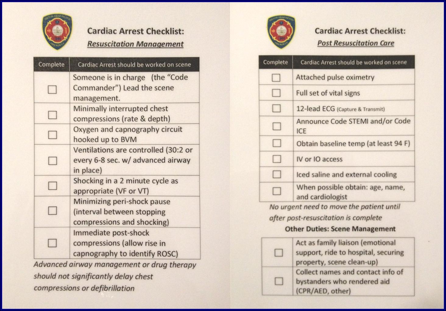
Cardiac Arrest Checklist

D: DYNAMIC PRIORITISATION, DECISION MAKING & DELEGATION

Do you have enough help? Does everyone have a task?

Based on your awareness and anticipation of the resuscitation be decisive on your immediate priorities, make and execute a plan of action, but remember to also be prepared to change as required.

Ensure you have enough resources available for everybody to be able to concentrate on their task fully and not be overloaded. As the resuscitation progresses it is likely your priorities will change and require you to adapt to these and redistribute the workload accordingly.

E: ERROR WISDOM & ENVIRONMENT

What are the potential errors that could occur? Can you carry out effective resuscitation within the environment the patient is in? What needs to change?

‘Forewarned is forearmed’

Be aware of the potential for errors. During stressful events such as resuscitation attempts, with a number of interventions performed and drugs administered, it is easy for these to occur. Considering this risk beforehand and utilizing tools such as checklists and pocket books can help minimize the potential for mistakes and refresh things in your head that you may not encounter very often in practice.

If the patient you are attending is in an unusual environment, or a scene that may pose a danger, consider the options you have to minimize hazards and allow you and the team to carry out the resuscitation safely. Even in domestic locations, during the initial stages of resuscitation it is easy to forget about the environment you are working in. Ensure you have enough space to work in and if not, move something!

I hope you have found this interesting. It may all seem like common sense and I am sure we all give these factors consideration every day with every incident we attend, however personally I feel having a structured approach is worth thinking about to reduce mental overload in times when we all need a little bit of extra brain capacity!

Reference

Logarajah, S. and Alinier, G. (2014). An Integrated ABCDE Approach To Managing Medical Emergencies Using CRM Principles (PDF – Subscription Required). Journal of Paramedic Practice, 6(12), pp.620-625.

Transcutaneous Pacing (TCP) With and Without Capture

EMS is dispatched to a private residence for 70-year-old female who is believed to be unconscious.

On arrival, the patient is found lying in bed unresponsive to painful stimuli. The patient’s skin is pale and clammy. Her shirt is damp. Snoring respirations are noted and a slow carotid pulse is present.

A medical history is obtained from family members and includes heart failure, stroke, and hypertension.

Vital signs are assessed.

  • RR: 8
  • HR: Less than 30
  • NIBP: 78/41
  • SpO2: Not registering
  • Temp: 96.1 F / 36.7 C
  • BGL: 101 mg/dL

The cardiac monitor is attached.

High degree AV block with wide complex escape rhythm.

The adult pads are placed and transcutaneous pacing is initiated.

The transcutaneous pacer is set for 70 PPM at 50 mA. Pacing spikes are visible with what appear to be large, corresponding QRS complexes.

The patient’s blood pressure improves slightly to 84/47 (confirmed by auscultation). However, paramedics are still concerned about the patient’s hypotension.

Additional treatments

  • IO access is obtained in right proximal tibia.
  • 0.5 mg of Atropine is administered x 3.
  • 9% normal saline is run wide open with an additional IV line established in the left lower extremity.

The patient begins to move and reaches for the pacing pads. However, she is still non-verbal and does not follow commands.

  • On arrival at the hospital the patient is transitioned to transvenous pacing.
  • She is sent to the cardiac cath lab where a permanent pacemaker is placed.
  • In the ICU the patient remains dangerously hypotensive in spite of dobutamine and levophed drips.

The patient eventually expires from multiple-system organ failure.

Discussion

Transcutaneous pacing (TCP) is a difficult skill that is often performed incorrectly. The problem of false capture (also known as echo distortion) is under-recognized and under-reported in the medical literature.

There are many reasons why medical professionals often fail to achieve true electrical and mechanical capture. Consider this excerpt from the Journal of Emergency Medicine where Douglas Ettin, M.D. and Thomas Cook, M.D. describe the difficulty.[1]

“Shortly after cardiac pacing was initiated, the patient’s systolic blood pressure dropped to 50 mmHg. The EKG monitor continued to demonstrate adequate ventricular capture by the pacer. The patient appeared to have palpable pulses; however, the rhythm contractions of the patient’s body from the pacer shocks made this assessment difficult. With the etiology of the patient’s hypotension unclear, the decision was made to use transthoracic ultrasonography to assess the adequacy of her ventricular contractions.”

“Initially, the ultrasound demonstrated ventricular contractions at a rate of 30-40 beats per minute. These heart contractions did not correspond with the surrounding thoracic muscle contractions generated by the pacer. The current was gradually increased to 110 mA, and the heart began to contract in unison with the pacer shocks. The patient’s blood pressure subsequently increased to 90 mmHg.”

You can see another example where an echo was used to verify capture here.

With false capture, you will generally see a near-vertical upstroke or down-stroke to the “phantom” QRS complex (which is actually electrical artifact created by the current passing between the pacing pads).

Classic “false capture” with near-vertical down-stroke of the (phantom) QRS complexes, slightly curved return to the isoelectric line, and unimpressive T-waves.

You will also note that the underlying rhythm can be seen in the absolute refractory period of one of the (presumed to be) paced QRS complexes (red circle). That is not scientifically possible!

In contrast, true electrical capture will show wide QRS complexes with tall, broad T-waves.

Transcutaneous pacing (TCP) with true electrical capture as evidenced by tall, broad T-waves. Capture was achieved at 110 mA (ems12lead.com).

Tips for success

  • Perform, but do not rely on a pulse check!
  • Use an instrument (SpO2, Doppler, capnography, or echo) to help confirm mechanical capture whenever possible
  • Do not be fooled by skeletal muscle contraction!
  • Know that the patient may become more alert whether capture is achieved or not
  • The most common reasons for “failure to capture” are insufficient milliamperes and poor pad placement!

More examples of transcutaneous pacing (TCP) with capture

Capture achieved at 140 mA and confirmed with sudden rise in ETCO2 (ems12lead.com).

110 mS

Capture achieved at 110 mA confirmed with SpO2 plethysmograph

Reference

  1. Ettin DCook T. Using ultrasound to determine external pacer capture. The Journal of Emergency Medicine. 1999;17(6):1007-1009.

Further reading

Pacing Artifact May Masquerade As Capture

This case was submitted by Roger Hancock with edits by Tom Bouthillet. Some details have been changed to protect patient confidentiality.

Everything You Should Know About Diltiazem (Cardizem)

A 50-year-old male with a history of hypertension (HTN) and atrial fibrillation (AF) presents to the Emergency Department with complaint of palpitations, which started while mowing the lawn.

He is alert and oriented with a Glasgow Coma Scale (GCS) of 15 and no signs of Hypoperfusion.

  • Heart Rate: 165/min, strong and irregular
  • Blood Pressure: 140/100 mmHg
  • Ventilatory Rate: 22/min
  • SpO2: 98% on room air

The patient is compliant with his medications and denies any allergies.

A 12 Lead ECG is recorded.

Atrial fibrillation with rapid ventricular response (RVR) and generalized ST-segment depression indicative of subendocardial ischemia.

The patient was treated with 20 mg of diltiazem (Cardizem) over 2 min, followed by 10 mg over 1 hr, and 0.25 mg digoxin (Lanoxin).

A rhythm change was noted on the monitor and another 12-lead ECG was recorded.

There is a sinus rhythm with left ventricular hypertrophy by limb lead voltage criteria and left atrial enlargement. There are no signs of subendocardial ischemia, suggesting the ST-segment depression was rate-related.

The patient was now asymptomatic and admitted for observation without further incident.

Understanding Diltiazem (Cardizem)

Diltiazem

Diltiazem (Cardizem) is a Class IV antiarrhythmic and one of the most common pharmacological agents used for treatment of AF with RVR.

Class IV antiarrhythmics are Calcium Channel Blockers (CCBs), which inhibit intracellular calcium influx via calcium channel antagonism. These particular pharmacological agents can be further divided into subdivisions based on their molecular composition:

Dihydropyridines (DHPs)

  • These CCBs can be easily identified by the last four letters of the generic name ending with “pine”.
  • DHP CCBs are more selective to peripheral vasculature than cardiac cells, leading to arterial smooth muscle relaxation and decreased Systemic Vascular Resistance (SVR), thus, decreasing afterload and Myocardial Oxygen Demand (MVO2).
  • Because of this peripheral calcium channel selectivity, they are commonly used for treatment of Hypertension and angina.
  • Their hemodynamic effects can be associated with adverse effects such as hypotension and reflex tachycardia secondary to sympathetic stimulation as a compensatory mechanism for the decreased cardiac output.

Examples include:

  • Amlodipine (Norvasc)
  • Nicardipine (Cardene)
  • Nifedipine (Procardia)

Non-dihydropyridines (NDHPs)

  • These CCBs are those which generic name does not end with “pine”.
  • Can be further divided into benzothiazepines (not to be confused with benzodiazepines) and phenylalkylamines.
  • Non-dihydropyridine CCBs are more selective to L-Type Calcium Channels in cardiac cells, such as the Sino Atrial Node (SAN) and Atrio Ventricular Node (AVN), although all CCBs cause peripheral vasodilation.
  • This Calcium Channel antagonism leads to decreased SAN chronotropic effect and decreased AVN conduction, making it useful for treatment of atrial arrhythmias such as AF, Atrial Flutter and Supra-ventricular Tachycardias (SVTs).

Examples include:

  • Benzothiazepines: Diltiazem
  • Phenylalkylamines: Verapamil

Vaughan-Williams Anti-arrhythmic Classification

There are four specific classes of antiarrhythmics with specific physiological functions divided into classes based on their mechanism of action. The rest of the pharmacological agents used as antiarrhythmics fall under the fifth class with different mechanisms of action from the previous classes.

One important aspect to understand is that although they are all antiarrhythmics, each class works under different mechanisms and therefore may have different effects on cardiac cells. Some target atrial, AV nodal or ventricular cells, while some have the capacity to address both atrial and ventricular arrhythmias.

Pharmacological Use

Diltiazem has a COR I, LOE-b classification, used for rate control of atrial arrhythmias, predominantly Atrial Fibrillation, and COR IIa, LOE-b for treatment of SVT with a reentry pathway mechanism.

2014 AHA/ACC/HRS Guideline for the Management of Patients With Atrial Fibrillation: Executive Summary: A Report of the American College of Cardiology/American Heart Association Task Force on Practice Guidelines and the Heart Rhythm Society. J Am Coll Cardiol. 2014;64(21):2246-2280. doi:10.1016/j.jacc.2014.03.021

2015 ACC/AHA/HRS Guideline for the Management of Adult Patients With Supraventricular Tachycardia: Executive Summary: A Report of the American College of Cardiology/American Heart Association Task Force on Clinical Practice Guidelines and the Heart Rhythm Society. J Am Coll Cardiol. 2015 Sep 16. pii: S0735-1097(15)06203-8. doi: 10.1016/j.jacc.2015.09.019.

Mechanism Of Action

  • Negative Chronotropic, Inotropic and Dromotropic effect by blocking L-Type Calcium Channels in cardiac tissue
  • Decreased Calcium influx affects Phase 2 of cardiac depolarization, delaying atrial and AVN conduction
  • This ultimately leads to decreased ventricular rate, with or without conversion to sinus rhythm

Caution

  • Diltiazem should be avoided in the presence of pre-excited AF with RVR, that is, AF in the presence of accessory pathway, i.e. Wolff Parkinson White (WPW) syndrome, as AVN blockage can lead to increased conduction through the accessory pathway, leading to life-threatening rapid ventricular rates.
  • Procainamide and Ibutilide are the preferred treatment of pre-excited AF with RVR and hemodynamically stable patients.
  • Diltiazem can be used in patients with AF and Heart Failure (HF) but with caution in reduced Left Ventricular Ejection Fraction and hypotension.

Dose and Administration

Although dosages may vary based on physician orders, protocols and age, a standard initial dose is 0.25 mg/kg, ranging between 10-20 mg over 2 minutes, with a second dose of 0.35 mg/kg, ranging between 20-25 mg over 2 minutes, often followed by a 5-10 mg/hr infusion.

Treatment of hemodynamically unstable patients in narrow QRS complex AF with RVR requires synchronized cardioversion at 120-200 J initially, and should not be delayed for administration of an anti-arrhythmic agent.

Conclusion

  • Diltiazem is a Class IV, non-dihydropyridine CCB anti-arrhythmic, serving as the most common pharmacological agent used for the treatment of AF and SVTs, for patients that are hemodynamically stable.
  • Caution should be used with CCBs and HF with decreased EF and hypotension.
  • Electrical Cardioversion should not be delayed for treatment with an anti-arrhythmic agent in the presence of Hypoperfusion and hemodynamically unstable patients.